
Discover what cryotherapy for skin is and how it effectively treats lesions and skin cancers. Get informed on this quick and safe procedure!
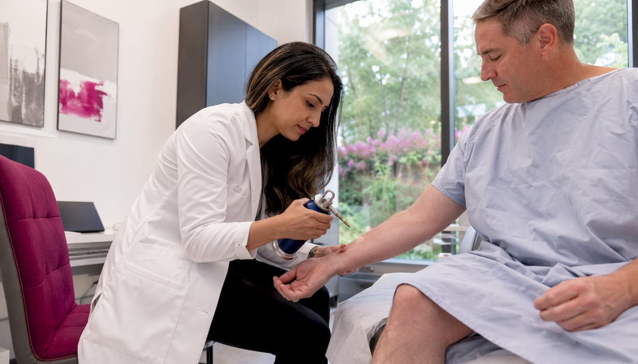
Cryotherapy for skin, also called cryosurgery or cryoablation, is a doctor-performed procedure that uses extreme cold, most often liquid nitrogen, to freeze and destroy abnormal skin tissue. Dermatologists use it routinely for three broad categories of conditions:
- Benign lesions: warts, skin tags, and seborrheic keratoses
- Premalignant lesions: actinic keratoses (rough, scaly patches caused by sun damage)
- Selected low-risk skin cancers: superficial basal cell carcinoma (BCC) and squamous cell carcinoma (SCC) in carefully chosen cases
The typical outcome is a quick office visit, usually under a minute for a small lesion, followed by expected redness, blistering, and scabbing that resolves over a few weeks, with good cosmetic results when the procedure is performed correctly.
Table of Contents
- What is cryotherapy for skin, and when do doctors use it?
- How does cryotherapy work, and what affects the results?
- What happens during a cryotherapy appointment?
- Aftercare and what the healing process actually looks like
- Benefits, risks, and who should not have cryotherapy
- When do dermatologists choose cryotherapy, and when do they recommend something else?
- Key Takeaways
- A dermatologist’s perspective on cryotherapy expectations
- Raodermatology’s approach to skin lesion evaluation and treatment
- Useful sources and further reading
- FAQ
What is cryotherapy for skin, and when do doctors use it?
The terms cryotherapy, cryosurgery, and cryoablation all describe the same basic concept: applying a cryogen cold enough to kill tissue. Liquid nitrogen is by far the most common agent, reaching temperatures around -196°C (-321°F). That extreme cold destroys cells faster and more reliably than older alternatives like carbon dioxide snow or dimethyl ether propane (DMEP) sprays, which are still occasionally used in primary care settings for minor benign lesions.
Cryosurgery is an outpatient procedure used across a wide spectrum of skin conditions, from the entirely harmless (a skin tag that catches on clothing) to the medically significant (an actinic keratosis that could progress to cancer if left untreated). For benign lesions, the goal is straightforward removal. For premalignant actinic keratoses, cryotherapy is one of the strongly recommended treatments in American Academy of Dermatology (AAD) guidelines, alongside topical agents and photodynamic therapy.
Where cryotherapy falls short is in cancer care beyond low-risk superficial tumors. It destroys tissue rather than preserving it, so a biopsy must come first whenever malignancy is suspected. It is also not appropriate for deep lesions, tumors with poorly defined margins, or any case where pathology is needed to confirm diagnosis and staging.
How does cryotherapy work, and what affects the results?
The freeze-thaw mechanism
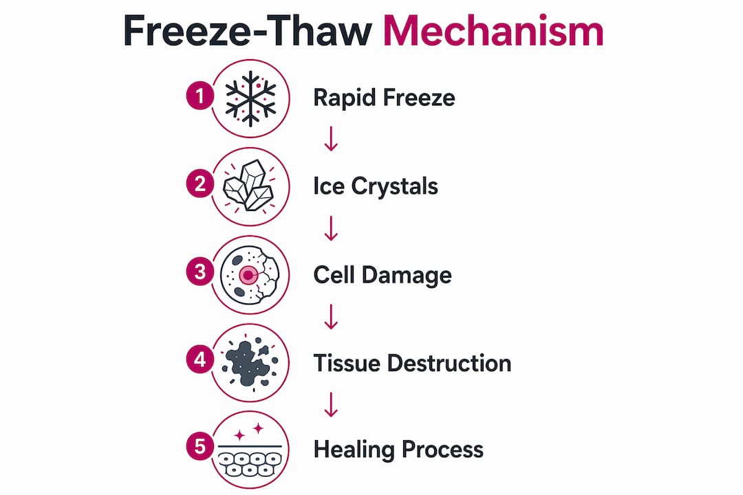
Rapid freezing causes ice crystals to form inside and between cells. That physical damage ruptures cell membranes. Simultaneously, the surrounding blood vessels go into spasm, cutting off circulation to the treated tissue. When the tissue thaws, a second wave of cellular injury occurs as osmotic pressure shifts and the vascular stasis resolves. Most practitioners use at least one complete freeze-thaw cycle; for malignant lesions, two or more cycles with a margin of 3–5 mm beyond the visible lesion edge are standard.
Delivery methods
| Method | How it works | Best for |
|---|---|---|
| Cryospray / cryogun | Liquid nitrogen sprayed directly onto lesion | Larger or irregular lesions |
| Cryoprobe | Metal tip cooled by liquid nitrogen, pressed to lesion | Precise, small lesions; eyelid margins |
| Cotton-tipped applicator | Cotton swab dipped in liquid nitrogen | Small benign lesions in primary care |
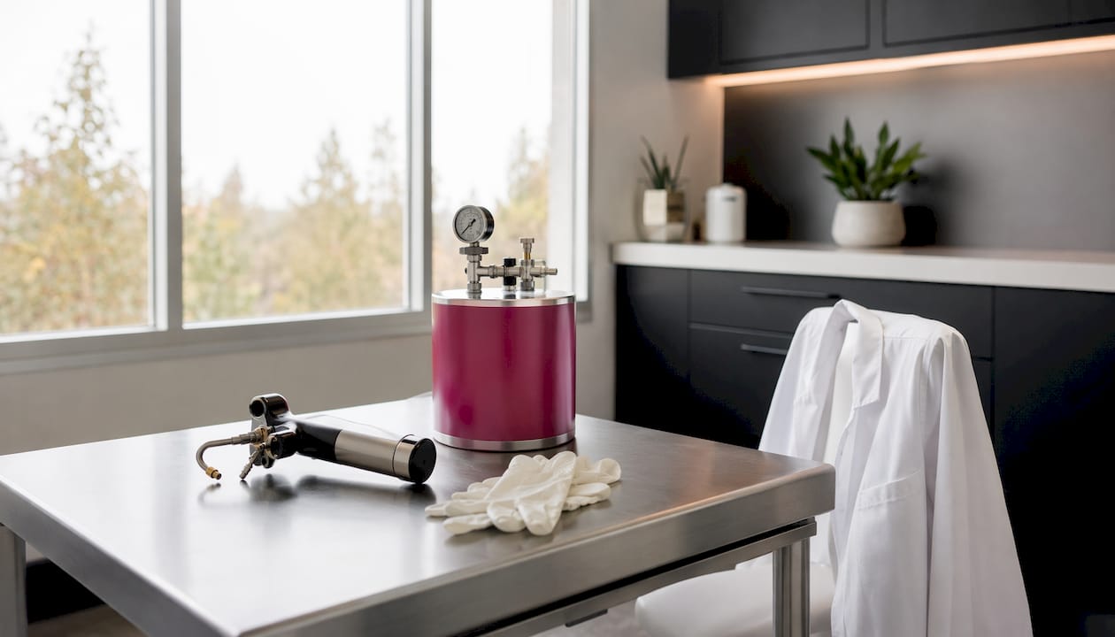
The spray method delivers the coldest, most consistent temperature and is the standard in most dermatology offices. Cotton applicators lose temperature quickly and are generally reserved for very superficial, small lesions.
Why freeze time changes everything
Freeze duration is the single most controllable variable in the procedure, and the data on actinic keratoses makes the point clearly:
| Freeze duration | Clearance rate (actinic keratoses) |
|---|---|
| Under 5 seconds | ~39% |
| 5–20 seconds | ~69% |
| Over 20 seconds | ~83% |
Source: NCBI Bookshelf / NBK554841
A longer freeze improves clearance but also increases the risk of scarring and pigment loss. That trade-off is exactly why operator skill determines outcomes. Spray angle, probe distance, and freeze duration all shift the depth and lateral spread of tissue destruction. Small errors compound.
Pro Tip: For thick or raised lesions, some dermatologists debulk the lesion with a curette before freezing. Removing the bulk first lets the cryogen penetrate more effectively, improving clearance without requiring an excessively long freeze.
What happens during a cryotherapy appointment?
Knowing the sequence of events before you walk in removes most of the anxiety. Here is how a typical visit unfolds:
- Lesion assessment. Your dermatologist examines the lesion visually and, if there is any concern about malignancy, discusses whether a biopsy is needed first. Cryotherapy destroys tissue architecture, so biopsy must precede freezing whenever cancer is suspected.
- Skin preparation. The area is cleaned. No shaving or special topical preparation is usually required for most lesions. If you take blood thinners or immunosuppressants, mention them at check-in, though they rarely change the plan for a simple benign lesion.
- The freeze. The clinician applies liquid nitrogen via spray, probe, or cotton applicator directly to the lesion and a small surrounding margin. For a small wart or skin tag, this takes seconds. Larger or thicker lesions may require 20–30 seconds or a repeat cycle. The entire in-office procedure is generally very brief for a single small lesion.
- Immediate sensation. Expect a sharp stinging or burning feeling during the freeze, followed by a dull ache for a few minutes afterward. Most patients tolerate this without injectable anesthetic. A topical numbing cream applied beforehand can reduce discomfort for sensitive areas.
- Post-procedure instructions. Before you leave, the clinician explains what the healing process will look like and what to watch for. Reviewing a dermatology appointment guide beforehand can help you prepare the right questions.
Aftercare and what the healing process actually looks like
The healing sequence after cryotherapy is predictable, but it looks alarming if you are not expecting it.
- Day 1: Redness and mild swelling at the treated site. This is normal.
- Days 1–3: A blister may form, sometimes filled with clear or blood-tinged fluid. Leave it intact. Puncturing the blister increases infection risk and can worsen scarring.
- Days 3–7: The blister flattens and a crust or scab forms.
- Weeks 1–3: The scab falls off on its own, revealing new skin underneath. Pigment changes (lighter or darker skin) may appear and often fade over several months.
- Sun protection: Apply broad-spectrum SPF 30+ to the healed area. UV exposure on newly formed skin worsens hyperpigmentation and delays full recovery.
Practical wound care is straightforward. Apply a thin layer of petroleum jelly (plain Vaseline works fine) to keep the area moist and reduce crusting. Avoid picking at the scab. For detailed aftercare steps, the cosmetic procedure aftercare guide at Raodermatology covers wound care and scar minimization in more depth.
Setting expectations about the healing cascade, the blistering, the scabbing, the temporary discoloration, significantly reduces unnecessary post-procedure calls and improves patient satisfaction, according to DermNet.
When to call your dermatologist: Contact the clinic if you notice spreading redness or red streaks (signs of infection), pus or foul-smelling drainage, fever, pain that worsens after day three rather than improving, or a treated area that shows no healing after three weeks.
Benefits, risks, and who should not have cryotherapy
The advantages
- Quick, office-based, no surgical incision required
- No injectable anesthetic needed for most lesions
- Low cost compared with excision or laser procedures
- Good cosmetic outcomes when technique is precise
Common side effects
- Pain and stinging during and immediately after the procedure
- Blistering and swelling (expected, not a complication)
- Temporary hyperpigmentation or hypopigmentation
- Scabbing lasting 1–3 weeks
Serious complications
Permanent hypopigmentation (a lighter patch of skin) is the most common lasting side effect and is more likely in patients with darker skin tones. Scarring can occur, particularly with overly aggressive freeze times. Rare nerve injury has been reported near digits and the periorbital area. For malignant lesions, recurrence rates treated with cryosurgery range from roughly 6% to 34%, so follow-up monitoring after treatment is important.
Contraindications
Cryotherapy is contraindicated in patients with cold-sensitive disorders, specifically:
- Raynaud disease (cold triggers vasospasm in fingers and toes)
- Cold urticaria (cold exposure causes hives or anaphylaxis)
- Cryoglobulinemia (abnormal proteins in the blood that precipitate in cold)
Patients with these conditions can experience serious systemic reactions from localized cryotherapy. Always disclose your full medical history before the procedure.
Pro Tip: Patients with darker skin tones (Fitzpatrick types IV–VI) face a higher risk of permanent hypopigmentation. A board-certified dermatologist should weigh this against the benefits and may recommend an alternative treatment for cosmetically sensitive areas.
When do dermatologists choose cryotherapy, and when do they recommend something else?
The decision depends on lesion type, number, depth, and whether pathology is needed.
| Situation | Preferred approach | Reason |
|---|---|---|
| Single actinic keratosis | Cryotherapy | Fast, effective, well-tolerated |
| Many actinic keratoses (field cancerization) | Field therapy (5-FU, imiquimod, PDT) | Treats visible and subclinical lesions across a broad area |
| Suspicious or undefined lesion margin | Biopsy first, then decide | Freezing destroys tissue needed for diagnosis |
| Low-risk superficial BCC/SCC | Cryotherapy (selected cases) | Appropriate with confirmed diagnosis and clear margins |
| High-risk or deep skin cancer | Surgical excision or Mohs | Requires margin control and pathology |
| Cosmetically sensitive area, darker skin | Laser or excision | Lower hypopigmentation risk |
AAD guidance is explicit: when a patient has numerous actinic keratoses, field-directed therapy is more practical and better tolerated than repeated spot cryotherapy treatments. Treating a dozen individual lesions one by one is painful, time-consuming, and misses the subclinical disease between visible spots.
For any lesion where cancer cannot be ruled out clinically, biopsy must come before cryotherapy. Freezing first and asking questions later is not acceptable practice at any reputable dermatology center.
Pro Tip: Ask your dermatologist specifically whether your lesion has been clinically confirmed as benign or premalignant before agreeing to cryotherapy. If there is any uncertainty, a biopsy is the right first step, not a freeze.
Key Takeaways
Cryotherapy is a fast, office-based procedure that effectively destroys benign and premalignant skin lesions when performed by a skilled dermatologist using the correct freeze duration and technique.
| Point | Details |
|---|---|
| Core definition | Cryotherapy uses liquid nitrogen to freeze and destroy abnormal skin tissue in an office setting. |
| Healing timeline | Expect blistering, scabbing, and full re-epithelialization over 1–3 weeks after treatment. |
| Freeze time matters | Clearance rates for actinic keratoses rise from 39% (under 5 seconds) to 83% (over 20 seconds). |
| Biopsy before freezing | Any lesion suspicious for cancer must be biopsied before cryotherapy, which destroys tissue needed for pathology. |
| Raodermatology | Raodermatology evaluates lesions across California, New Jersey, and New York and determines whether cryotherapy or an alternative treatment is appropriate for each patient. |
A dermatologist’s perspective on cryotherapy expectations
Cryotherapy gets undersold and oversold at the same time, and that gap is where patient frustration lives. Patients sometimes arrive expecting a painless, invisible fix. Others have read horror stories about scarring and refuse a procedure that would genuinely help them. Neither picture is accurate.
The honest version: cryotherapy is one of the most useful tools in a dermatologist’s office precisely because it is fast, inexpensive, and does not require a surgical suite. But “simple” does not mean “automatic.” The difference between a clean cosmetic result and a permanent white patch often comes down to seconds of freeze time and a millimeter of margin. That is not a reason to avoid the procedure. It is a reason to have it done by someone who performs it regularly and knows your skin type.
The other thing patients consistently underestimate is the healing process. The blister that forms the next day looks far worse than it is. Leaving it alone, keeping it moist, and protecting it from the sun is genuinely all most people need to do. The patients who pick at the scab or skip sunscreen are the ones who end up with the outcomes they were trying to avoid.
Shared decision-making matters here more than in many procedures. The right treatment for an actinic keratosis on a fair-skinned patient with one lesion is not automatically the right treatment for a patient with twenty lesions and a Fitzpatrick type V complexion. A good dermatologist walks through those variables with you before picking up the spray canister.
Raodermatology’s approach to skin lesion evaluation and treatment
Raodermatology’s medical dermatology teams across California, New Jersey, and New York evaluate skin lesions with the same starting point every time: confirm what you are treating before you treat it. For patients with warts, skin tags, actinic keratoses, or lesions that concern them, that means a thorough clinical evaluation, biopsy when indicated, and a treatment plan built around the specific lesion, skin type, and cosmetic priorities.
Cryotherapy is one of several skin cancer and lesion treatment options available at Raodermatology, alongside surgical excision, photodynamic therapy, and topical field treatments. If you have a lesion you want evaluated, schedule a consultation at one of Raodermatology’s locations. The right treatment starts with the right diagnosis.
Useful sources and further reading
The following authoritative sources provide the clinical guidelines and procedural detail referenced throughout this article:
- Cutaneous Cryosurgery — NCBI Bookshelf (NBK554841): Comprehensive StatPearls review of cryosurgery indications, technique, and outcomes.
- Cryotherapy in Dermatology — StatPearls (NBK482319): Detailed technical review covering freeze-thaw cycles, margins, and operator variables.
- Cryotherapy for the skin — MedlinePlus Medical Encyclopedia: Patient-facing overview from the U.S. National Library of Medicine.
- Actinic Keratosis: Diagnosis and Treatment — American Academy of Dermatology: AAD public guidance on treatment options including cryotherapy and field therapies.
- Cutaneous Cryosurgery for Common Skin Conditions — AAFP/AFP: Clinical practice article covering indications, biopsy requirements, and technique.
- Cryotherapy — DermNet: Accessible clinical reference covering contraindications, complications, and aftercare.
Consult these resources or speak directly with a board-certified dermatologist for guidance specific to your situation. This article provides general medical information, not personalized medical advice.
FAQ
What does cryotherapy actually feel like during the procedure?
Most patients describe a sharp stinging or burning sensation during the freeze, followed by a dull ache for a few minutes. Injectable anesthetic is not usually required, though a topical numbing cream can be applied beforehand for sensitive areas.
Is cryotherapy safe for children?
Cryotherapy can be effective in children, but comparative studies on molluscum contagiosum found it causes more pain, blistering, and superficial scarring than topical alternatives. Pediatric dermatologists often prefer topical treatments first and reserve cryotherapy for cases that do not respond.
How many cryotherapy sessions will I need?
Many benign lesions clear in one session. Warts and thicker lesions often require two or more treatments spaced several weeks apart. Your dermatologist will assess the lesion’s response at a follow-up visit before deciding whether retreatment is needed.
Can cryotherapy remove skin cancer?
Cryotherapy can treat selected low-risk superficial skin cancers, but it is not appropriate for most malignant lesions. Recurrence rates for malignant lesions treated with cryosurgery range from roughly 6% to 34%, and a confirmed biopsy diagnosis is required before freezing. Surgical excision or Mohs surgery is the standard of care for most skin cancers.
Will cryotherapy leave a scar or white spot?
Temporary pigment changes are common and usually fade over several months. Permanent hypopigmentation (a lighter patch) is the most common lasting side effect and is more likely with longer freeze times or in patients with darker skin tones. Precise technique by an experienced dermatologist significantly reduces this risk.
Recommended
Filed under: