
Discover what a skin lesion is, its types, causes, and essential treatments. Stay informed for better skin health!

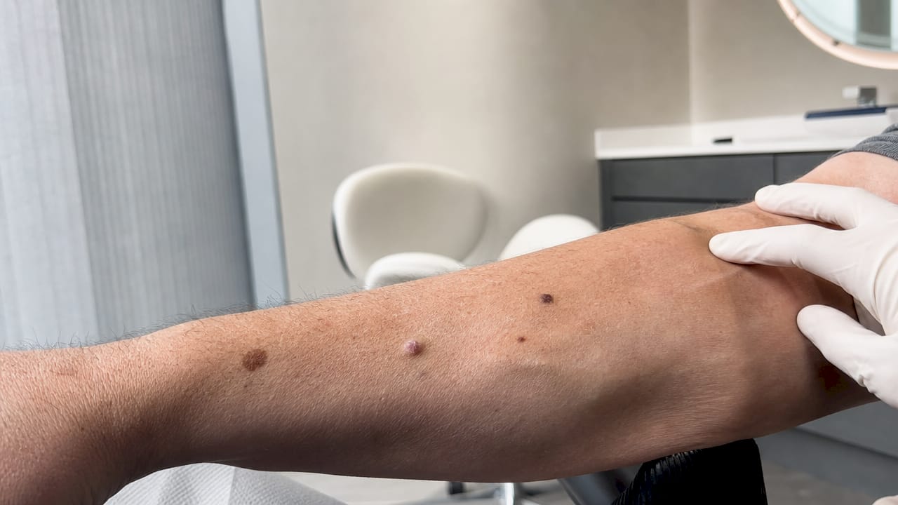
A skin lesion is defined as any distinct area of skin that differs from the surrounding tissue in color, shape, size, or texture, and can range from a completely harmless mole to a malignant melanoma. Most people notice a new spot or patch and immediately wonder whether it signals something serious. The honest answer is that most lesions are benign, but visual similarity between harmless and dangerous growths makes professional evaluation non-negotiable. Causes span everything from viral infections and sun damage to systemic diseases and genetic predisposition. Recognizing what you are looking at, and knowing when to act, is the foundation of smart skin health.
What is a skin lesion, and how are they classified?
A skin lesion is any spot differing from surrounding skin by color, shape, size, or texture, whether caused by localized damage or a systemic disorder. Dermatologists classify lesions in two broad ways: by their clinical behavior (benign, premalignant, or malignant) and by their physical morphology, meaning the structural form the lesion takes.
Morphology is the language clinicians use to describe what they see before ordering any test. The core terms every patient should recognize are:
- Macule: A flat, colored spot under 1 cm, such as a freckle.
- Papule: A raised, solid bump under 1 cm, like a small wart.
- Plaque: A raised, flat-topped area larger than 1 cm, common in psoriasis.
- Nodule: A deeper, firmer lump extending into the dermis, such as a cyst.
- Vesicle/Bulla: A fluid-filled blister, small or large, seen in conditions like shingles.
Beyond morphology, lesions split into pigmented and non-pigmented categories. Pigmented lesions, including moles and seborrheic keratoses, contain melanin. Non-pigmented lesions, such as warts or basal cell carcinomas, do not. Seborrheic keratoses appear stuck-on and can closely mimic premalignant actinic keratoses, but they carry zero cancer risk. That distinction matters because the two conditions look nearly identical to an untrained eye.
| Lesion type | Behavior | Common examples |
|---|---|---|
| Benign | Non-cancerous, stable | Moles, seborrheic keratoses, warts |
| Premalignant | May progress to cancer | Actinic keratoses, dysplastic nevi |
| Malignant | Cancerous, requires treatment | Melanoma, basal cell carcinoma, squamous cell carcinoma |
Pro Tip: Photograph any new or changing spot with your phone every 4 to 6 weeks. A side-by-side comparison over time is often the clearest early warning signal you can bring to a dermatologist.
What causes skin lesions and which factors raise cancer risk?
Understanding the cause of a lesion is as important as its appearance, because the underlying driver determines both the risk level and the correct treatment path. Causes fall into four main categories: infectious, inflammatory, genetic, and neoplastic.
Infectious causes include the human papillomavirus (HPV), which produces common warts, and herpes zoster, which produces the painful blistering rash of shingles. Bacterial infections cause impetigo and folliculitis. Fungal infections produce ringworm and tinea versicolor, both of which alter skin pigmentation. Inflammatory causes include allergic contact dermatitis, eczema, and psoriasis, all of which create plaques or patches that can be mistaken for more serious lesions. Genetic factors drive conditions like neurofibromatosis, where multiple benign tumors develop across the skin. Neoplastic causes represent abnormal cell growth, which is where malignancy risk enters the picture.
Several factors specifically increase the risk that a lesion will become or already is malignant:
- Cumulative UV exposure. Sun exposure drives both premalignant actinic keratoses and skin cancers. Sun protection prevents actinic keratoses and reduces long-term cancer risk.
- Immunosuppression. Patients on organ transplant medications or those with HIV face significantly higher rates of skin cancer because immune surveillance is reduced.
- Fair skin and family history. Both lower the threshold at which UV damage becomes dangerous.
- Lesion evolution. A mole that changes shape, bleeds, or becomes tender is a stronger warning sign than its size alone.
The ABCDE criteria give you a structured way to assess any pigmented lesion at home. Melanoma risk is evaluated by checking for Asymmetry, Border irregularity, Color variation, Diameter over 6 mm, and Evolution over time. Evolution, meaning any new change in an adult, is the single most critical warning sign. A small melanoma that lacks large size can still be dangerous if it starts bleeding or becomes tender.
How are skin lesions diagnosed by dermatologists?
Diagnosis relies primarily on clinical examination, combining visual inspection with tactile assessment of texture, firmness, and depth. Dermatologists first categorize a lesion as infection-related, inflammatory, pigmented, or a growth or tumor type. That initial classification determines which diagnostic tools come next.
Dermatoscopy is the most significant advancement in non-invasive lesion evaluation available today. A dermatoscopy guide explains how this handheld device uses polarized light to reveal subsurface structures invisible to the naked eye, including vascular patterns and pigment networks that distinguish benign nevi from early melanomas. Dermatoscopy reduces unnecessary biopsies and increases diagnostic accuracy.
When a lesion remains uncertain after visual and dermatoscopic examination, biopsy is the definitive next step. The procedure involves removing a small tissue sample, either by shave, punch, or excisional technique, and sending it to a dermatopathology lab for microscopic analysis. Dermatopathology is the subspecialty that examines skin tissue at the cellular level, and it provides the only definitive answer on whether a lesion is malignant.
Medical history plays a larger role than most patients expect. A dermatologist will ask how long the lesion has been present, whether it has changed, and whether you have a personal or family history of skin cancer. Lesion morphology and patient history together guide every clinical decision, from watchful waiting to urgent excision.
Pro Tip: Before your appointment, write down when you first noticed the lesion, any changes you have observed, and any medications you take. Dermatologists make faster and more accurate assessments when this information is ready upfront.
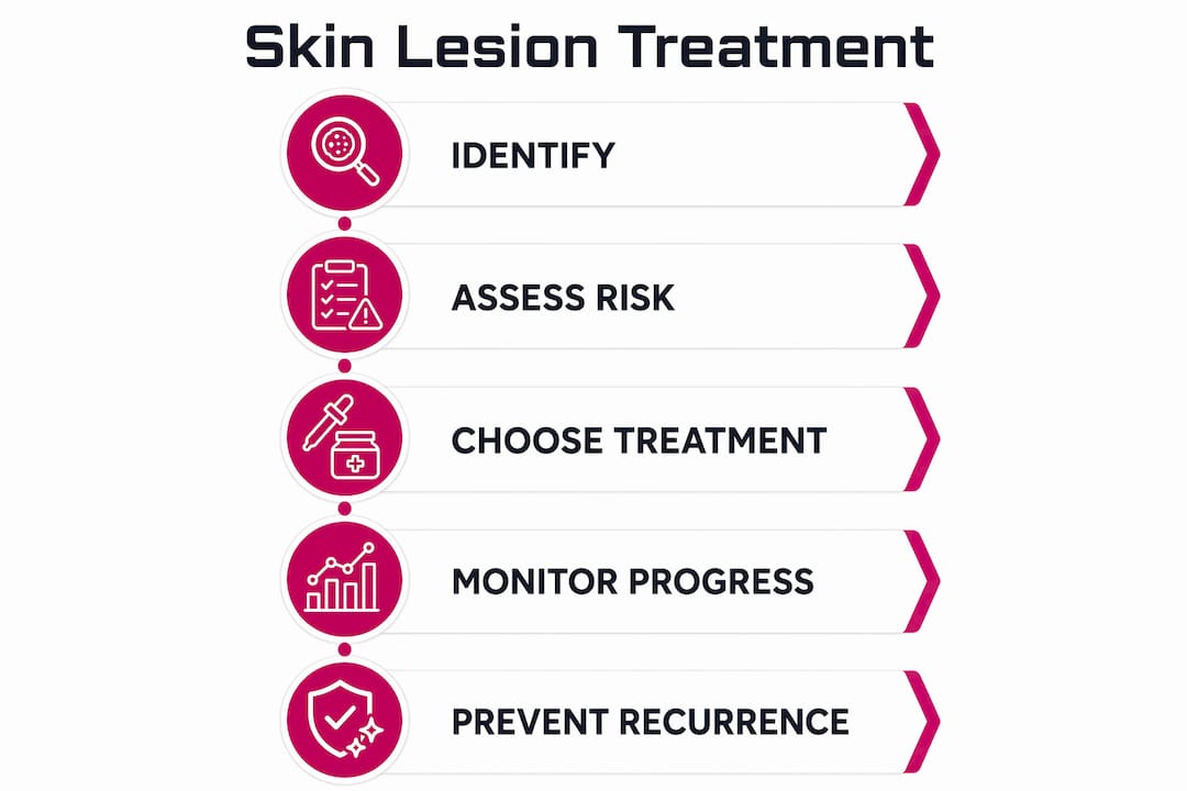
What treatment options exist for skin lesions?
Treatment depends entirely on the lesion type, its malignant potential, its location, and the patient’s preferences. Treatment choice reflects lesion type, location, and individual factors, which is why a one-size-fits-all approach does not work in dermatology.
Benign lesions that are asymptomatic and stable often require no treatment at all. When removal is desired for cosmetic reasons or because a lesion catches on clothing, dermatologists use cryotherapy (liquid nitrogen freezing), laser therapy, electrocautery, or simple shave excision. Laser therapy options have expanded considerably, with pulsed dye lasers targeting vascular lesions and fractional CO2 lasers addressing pigmented or textural irregularities.
Premalignant lesions, particularly actinic keratoses, require a more strategic approach. Actinic keratoses carry a progression risk to squamous cell carcinoma ranging from less than 1% to 10% per lesion per year. Treatment balances lesion-directed methods, such as cryotherapy or curettage on individual spots, with field-directed therapies that treat the broader sun-damaged area. Field therapy matters because subclinical damage extends well beyond visible lesions. Topical agents like 5-fluorouracil cream and photodynamic therapy address this wider zone of risk. Solar keratoses also signal increased future skin cancer risk, prompting enhanced surveillance even after treatment.
Malignant lesions require surgical excision with clear margins as the standard of care. Mohs micrographic surgery is used for high-risk locations like the face, where tissue preservation matters. Follow-up schedules after malignant lesion removal are non-negotiable, since recurrence and new primary cancers are real risks.
Across all lesion types, sun protection is the most cost-effective prevention strategy available. Daily broad-spectrum SPF 30 or higher, protective clothing, and avoiding peak UV hours reduce both new lesion formation and the progression of existing premalignant spots.
| Treatment approach | Best suited for | Method examples |
|---|---|---|
| Watchful waiting | Stable benign lesions | Periodic monitoring, photography |
| Lesion-directed removal | Individual benign or premalignant spots | Cryotherapy, laser, shave excision |
| Field-directed therapy | Sun-damaged skin with multiple actinic keratoses | 5-fluorouracil cream, photodynamic therapy |
| Surgical excision | Malignant or high-risk lesions | Standard excision, Mohs surgery |
Key takeaways
Accurate identification of a skin lesion’s type, cause, and risk level is the single most important factor in choosing the right treatment and avoiding delayed cancer diagnosis.
| Point | Details |
|---|---|
| Definition matters | A lesion is any skin area differing in color, shape, size, or texture from surrounding skin. |
| Most are benign | The majority of skin growths are harmless, but professional evaluation rules out malignancy. |
| ABCDE criteria save lives | Asymmetry, border, color, diameter, and evolution guide early melanoma detection at home. |
| Actinic keratoses need treatment | These premalignant spots carry real cancer progression risk and require lesion or field therapy. |
| Sun protection is prevention | Daily SPF use reduces new lesion formation and slows progression of existing premalignant spots. |
What I’ve learned from watching patients wait too long
In my experience reviewing dermatology cases, the pattern that concerns me most is not the lesion itself. It is the delay. Patients routinely wait six months to two years before having a changing spot evaluated, often because they assume it is “just a mole” or because they do not want to seem alarmist. That hesitation is where outcomes diverge.
The ABCDE criteria are genuinely useful for self-monitoring, but they have a real limitation: evolution is the most important criterion, and you cannot assess evolution from a single observation. You need a baseline. I would argue that every adult should photograph their full skin surface at least once a year, not because most spots are dangerous, but because having a reference point transforms a vague concern into a concrete, answerable question at your next dermatology visit.
Advances in dermatoscopy and dermatopathology have made diagnosis faster and less invasive than it was even a decade ago. Minimally invasive removal methods mean that treating a benign lesion cosmetically, or excising a premalignant one early, carries very little burden. The calculus has shifted. Waiting is now the higher-risk choice. Most lesions will turn out to be nothing. But the ones that are not something are caught earliest by people who act promptly and see a qualified dermatologist regularly.
— Krunal
Expert skin lesion care at Raodermatology
Raodermatology, founded by Dr. Babar K. Rao with over 25 years of clinical experience across California, New Jersey, and New York, provides full-spectrum skin lesion evaluation and treatment under one roof.

From dermatoscopic examination and biopsy to cryotherapy, laser removal, and surgical excision, the practice handles benign, premalignant, and malignant lesions with individualized care plans. The team’s skin cancer services include early detection screenings, actinic keratosis management, and Mohs surgery referral coordination. If you have noticed a new or changing spot, the most productive next step is a professional evaluation. Schedule a consultation with Raodermatology and get a clear, expert answer about what you are dealing with.
FAQ
What is a skin lesion in simple terms?
A skin lesion is any spot, patch, bump, or sore on the skin that looks or feels different from the surrounding skin. Lesions can be flat or raised, pigmented or colorless, and range from harmless to cancerous.
Are all skin lesions cancerous?
No. Most skin growths are benign and pose no cancer risk. However, some lesions are premalignant or malignant, which is why clinical evaluation is recommended for any new or changing spot.
What does a skin lesion look like?
Appearance varies widely by type. A macule is flat and discolored, a papule is a small raised bump, and a nodule is a deeper lump. Using skin lesion pictures alongside the ABCDE criteria helps identify warning signs, but a dermatologist’s assessment is the only reliable confirmation.
When should I see a doctor about a skin lesion?
See a dermatologist if a lesion changes in size, shape, or color, bleeds without injury, itches persistently, or is new in adulthood. Evolution of features is the strongest early warning sign for malignancy.
How are skin lesions treated?
Treatment depends on the lesion type. Benign lesions may be monitored or removed with cryotherapy, laser, or excision. Premalignant lesions like actinic keratoses require lesion-directed or field-directed therapy. Malignant lesions are treated with surgical excision, often followed by structured monitoring.
Recommended
- Types of skin cancer: Risks, signs, and prevention explained | Rao Dermatology
- Common Causes of Skin Cysts: Types, Symptoms & Treatment Options | Rao Dermatology
- Understanding Bacterial Skin Infections: Causes, Types & Treatment Options | Rao Dermatology
- What Are Cysts? Causes, Symptoms, and Treatment Options | Rao Dermatology
Filed under: