
Learn how dermatopathology works, why it matters for accurate skin diagnosis, and how to use this knowledge to get better care from your dermatologist.
Most people assume a dermatologist diagnoses skin conditions the same way a mechanic spots a dent, just by looking. But many of the most critical skin health decisions rely on what happens in a laboratory, not a waiting room. Dermatopathology is the medical specialty that examines skin tissue under a microscope to identify diseases with precision that no visual exam alone can match. Understanding how it works puts you in a much stronger position when talking with your doctor, especially when the stakes involve skin cancer, unusual rashes, or complex inflammatory conditions.
Table of Contents
- What is dermatopathology and why does it matter?
- How dermatopathologists diagnose skin diseases
- Pitfalls, limitations, and why clinical context matters
- Emerging innovations: The future of dermatopathology
- The real value of dermatopathology: What most patients never realize
- How Rao Dermatology supports your skin health with expert dermatopathology
- Frequently asked questions
Key Takeaways
| Point | Details |
|---|---|
| Definition clarified | Dermatopathology is the microscopic study of skin diseases using biopsies, crucial for accurate diagnosis. |
| Essential for tough cases | It’s essential when visual exams cannot distinguish between similar-looking rashes or tumors. |
| Advanced tools improve accuracy | AI and advanced methods now deliver up to 90% diagnostic accuracy but expert review remains critical. |
| Patient empowerment | Understanding dermatopathology helps you ask better questions and get more precise care. |
What is dermatopathology and why does it matter?
Dermatopathology sits at the crossroads of two specialties: dermatology and pathology. It focuses on diagnosing diseases of the skin, hair, and nails by analyzing tissue samples at the cellular and molecular level. The doctor who does this work is called a dermatopathologist, a physician with specialized training in both fields.
Here is how the three roles break down:
- Dermatologist: Examines your skin clinically, identifies visible signs of disease, and performs biopsies.
- Pathologist: Analyzes tissue samples from many organ systems but may not specialize in skin.
- Dermatopathologist: Focuses exclusively on skin tissue, combining deep clinical and lab expertise.
For patients in California, New Jersey, and New York, dermatopathology consultations most often come up when:
- A mole or growth looks suspicious during a routine exam.
- An unexplained rash does not respond to standard treatment.
- A skin infection resists typical therapies.
- Conditions in children or teens require specialized analysis through pediatric dermatology.
Skin biopsies are the foundation of dermatopathology. A small piece of skin is removed and sent to a lab, where microscopic examination of skin biopsies using hematoxylin and eosin (H&E) staining reveals the cellular structure in detail. Advanced molecular tests add another layer of precision when standard stains are not enough.
AI-assisted tools have pushed diagnostic accuracy to nearly 90% in recent research, but these tools still work best alongside expert human review.
Pro Tip: Before any biopsy, ask your board-certified dermatologist whether the sample will be reviewed by a dedicated dermatopathologist. Not all labs provide that level of specialization.
How dermatopathologists diagnose skin diseases
The path from a skin concern to a confirmed diagnosis follows a clear sequence. Here is what typically happens:
- Clinical skin exam: Your dermatologist identifies a lesion or abnormality that needs further evaluation.
- Biopsy collection: A sample of skin tissue is removed using a punch, shave, or excisional technique.
- Lab processing: The tissue is fixed, embedded in paraffin, sliced thin, and mounted on glass slides.
- Microscopic analysis: The dermatopathologist examines the slides using H&E staining to assess cell shape, arrangement, and structure.
- Special stains and advanced testing: Additional techniques are applied based on what the initial review shows.
- Diagnostic report: A written report is sent to your treating physician with findings, interpretation, and diagnosis.
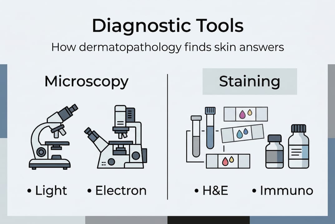
The core diagnostic methods include H&E staining, light microscopy, electron microscopy, fluorescence microscopy, immunohistochemistry (IHC), and molecular diagnostics including next-generation sequencing (NGS). Each tool is suited to different conditions.
| Test type | Best used for |
|---|---|
| H&E staining | Rashes, inflammatory conditions, common tumors |
| Immunohistochemistry (IHC) | Melanoma, basal cell carcinoma, lymphoma |
| Molecular/NGS testing | Rare genetic conditions, atypical pigmented lesions |
| Fluorescence microscopy | Autoimmune blistering diseases |
For conditions like psoriasis vs. eczema, these tests help clinicians distinguish between conditions that look nearly identical on the surface. And for melanoma detection, IHC and molecular tools can be the difference between a correct diagnosis and a dangerous miss.
Pro Tip: If you have a pigmented lesion or a rare skin condition, ask specifically whether IHC or molecular testing will be part of your biopsy workup. You have every right to understand what tests are being run.
Pitfalls, limitations, and why clinical context matters
Dermatopathology is precise, but it is not infallible. Several factors can make diagnosis genuinely difficult, and patients should understand why.

As research notes, inflammatory diseases often share patterns, and distinguishing benign from malignant neoplasms requires careful clinical input alongside the lab findings. This means a tissue sample alone does not always tell the full story.
Factors that increase diagnostic complexity include:
- Similar microscopic patterns: Some inflammatory conditions like psoriasis and psoriasis and dermatitis overlap significantly under the microscope.
- Prior treatments: Medications, steroids, or previous biopsies can alter tissue appearance and mask true findings.
- Rare variants: Uncommon presentations of known diseases do not always match textbook patterns.
- Incomplete clinical history: Missing information about medications, sun exposure, or prior diagnoses can lead a dermatopathologist astray.
Here is a quick comparison of what separates benign from malignant findings in common biopsies:
| Feature | Benign | Malignant |
|---|---|---|
| Cell size and shape | Uniform, regular | Variable, irregular |
| Nuclear appearance | Small, round nuclei | Large, atypical nuclei |
| Mitosis (cell division) | Rare | Frequent, abnormal |
| Tissue architecture | Organized layers | Disrupted, invasive growth |
A WebMD overview reinforces that a dermatopathologist’s role goes well beyond reading slides. They serve as a diagnostic partner to your clinical team, correlating what they see in the lab with what your dermatologist reports from your exam.
This clinicopathologic correlation is where errors are caught and accurate diagnoses are confirmed. If your clinical history is incomplete when your biopsy is submitted, the analysis could miss critical context. Always give your care team the full picture, including medications, travel history, and any relevant family history of skin cancer.
Emerging innovations: The future of dermatopathology
The field is evolving quickly, and some of the changes directly affect how accurately and efficiently you could receive a diagnosis.
AI models like DermpathNet and HistoGPT are now approaching 90% diagnostic accuracy in controlled studies, making them powerful screening tools. Digital pathology, where slides are scanned and reviewed virtually, is removing geographic barriers and allowing expert consultation across time zones.
Other notable advances include:
- Reflectance confocal microscopy: A non-invasive imaging tool that allows real-time examination of skin at near-cellular resolution, reducing the need for some biopsies.
- Next-generation sequencing (NGS): Identifies genetic mutations in tumor cells with high precision, critical for rare or aggressive skin cancers.
- Whole slide imaging (WSI): Digitizes entire tissue slides for remote collaboration and AI-assisted review.
- HistoGPT: A language model trained on histopathology data that generates diagnostic interpretations similar to a specialist’s report.
According to emerging technique research, tools like HistoGPT and reflectance confocal microscopy set new diagnostic standards but still require standardization before replacing human review entirely.
For patients, these advances mean faster turnaround times, more precise results, and better outcomes for advanced melanoma care and conditions treated with innovative approaches like photodynamic therapy. But the core message stays the same: technology enhances the expert, it does not replace them.
The real value of dermatopathology: What most patients never realize
Here is something worth saying directly: most patients never meet their dermatopathologist. You go to your appointment, have a biopsy, and wait for results. Someone you have never seen, in a lab you have never visited, makes one of the most important calls about your health.
That quiet dynamic is exactly why patient engagement matters. The clinicopathologic team, your dermatologist working in tandem with a dermatopathologist, consistently outperforms any single clinician working alone. This is not just a workflow preference. It is a structural advantage that reduces misdiagnosis.
From our experience at Rao Dermatology, patients who ask questions get better care. Not because asking changes the science, but because it ensures the full clinical picture reaches the lab. The more your care team knows, the more accurate the analysis becomes.
We also believe second opinions have enormous value, particularly for ambiguous or rare findings. Pathology reports are not always final verdicts. They are expert interpretations, and a second expert may see something different.
Working with board-certified dermatologists who prioritize integrated care gives you access to both sides of that equation.
Pro Tip: Request a copy of your pathology report after every biopsy. Ask your provider to walk you through what it says. You deserve to understand the findings, not just the conclusion.
How Rao Dermatology supports your skin health with expert dermatopathology
At Rao Dermatology, we integrate clinical expertise with a deep commitment to diagnostic accuracy across our locations in California, New Jersey, and New York. Dr. Babar K. Rao and our team bring over 25 years of experience to both the exam room and the evaluation process.

Whether you need evaluation of a suspicious lesion, management of a complex inflammatory condition, or access to our full range of dermatopathology services, we connect the clinical and laboratory sides of your care. Our skin cancer care programs and medical dermatology services reflect that integrated approach. Reach out to schedule a consultation and experience the difference that informed, coordinated care makes.
Frequently asked questions
What does a dermatopathologist do?
A dermatopathologist is a medical doctor who identifies and explains skin diseases by examining skin samples under a microscope. They use microscopic and molecular tools to deliver precise diagnoses that guide your treatment plan.
When is a dermatopathology report especially important for skin health?
A dermatopathology report is most critical when a visual exam cannot confirm a diagnosis, such as with unexplained rashes, skin tumors, or suspected cancer. Microscopic exam provides the cellular-level clarity that clinical observation alone cannot.
How accurate are dermatopathology diagnoses?
Modern dermatopathology achieves up to 90% accuracy when AI-assisted tools are combined with expert human review. Accuracy is highest when full clinical history accompanies the tissue sample.
Can I request my biopsy to be reviewed by a dermatopathologist?
Yes, absolutely. You can and should request specialist review, particularly for serious, rare, or unclear skin conditions. Expert review remains the consensus gold standard in current dermatopathology practice.
Recommended
- Dermatopathology | Rao Dermatology
- Why Choose a Board-Certified Dermatologist: Benefits and Expertise Explained | Rao Dermatology
- National Healthy Skin Month: Essential Dermatologist-Approved Tips for Year-Round Skin Health | Rao Dermatology
- Essential Guide to Skin Self-Examination: What to Look for Between Dermatology Visits | Rao Dermatology
- Was ist ein Pathologiebefund? 30% sind gutartig erklärt
Filed under: