
Discover effective ways to treat post-inflammatory hyperpigmentation and learn how to speed up recovery with proper care and sunscreen.
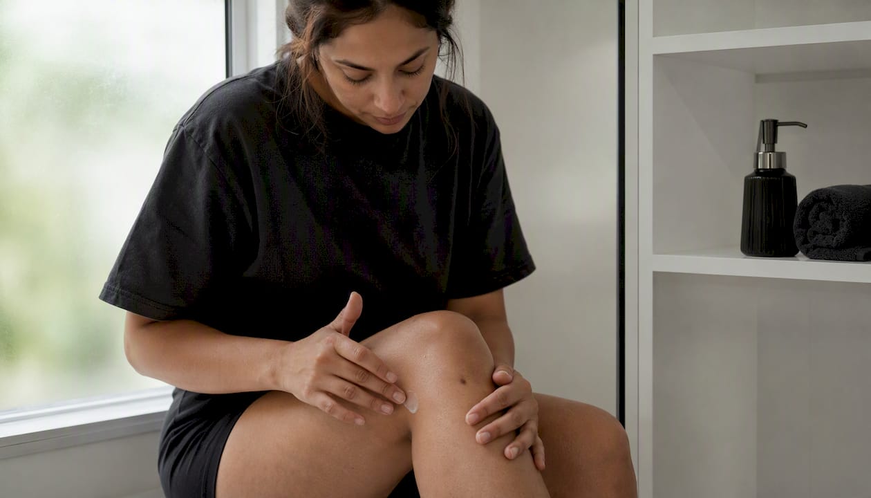
Post-inflammatory hyperpigmentation (PIH) is the darkening of skin that follows inflammation or injury, caused by excess melanin deposited in the epidermis or dermis. It is not a disease itself but a predictable skin response, and the single most important thing you can do right now is apply a broad-spectrum sunscreen every morning and stop touching or picking at affected areas.
Immediate dos and don’ts:
- Do apply broad-spectrum sunscreen daily, even indoors near windows
- Do treat active acne or eczema promptly to cut off the inflammatory signal
- Don’t pick, squeeze, or scrub affected skin, even gently
Timeline at a glance: Epidermal PIH (the more common, brown/tan variety) often fades over several months with consistent care. Dermal PIH, which sits deeper and appears blue-gray, can take years and sometimes does not fully resolve.
Pro Tip: Start sunscreen the same day you notice a new dark spot forming, not after the inflammation clears. Exposure to UV after injury is one of the fastest ways to lock in pigment that would otherwise fade on its own.
If spots are spreading rapidly, have irregular borders, or have persisted beyond three to four months without any improvement, schedule a dermatology evaluation rather than continuing to self-treat.
Key Takeaways
Post-inflammatory hyperpigmentation is predictable, treatable, and largely preventable when photoprotection and inflammation control are prioritized from the start.
| Point | Details |
|---|---|
| Treat inflammation first | Lightening agents work poorly against active acne or eczema; control the trigger before starting topicals. |
| Sunscreen is non-negotiable | Daily broad-spectrum SPF 30–60 prevents worsening and recurrence, even after spots have cleared. |
| Epidermal PIH timeline | Brown/tan spots often improve within 6–12 months with consistent topical therapy and photoprotection. |
| Combination therapy wins | Hydroquinone plus a retinoid, with or without a short-term steroid (≤8 weeks), outperforms monotherapy. |
| Rao Dermatology | Offers structured PIH evaluation, prescription topical management, and procedural options across California, New Jersey, and New York. |
Table of Contents
- What is post-inflammatory hyperpigmentation, and how is it different from other dark spots?
- Common causes and triggers of PIH
- Who is most at risk for PIH?
- How inflammation leads to dark spots: the mechanism behind PIH
- How PIH looks on the skin and what clinicians examine
- How dermatologists evaluate and diagnose PIH
- The stepwise approach to treating PIH
- Topical treatments for PIH: what works and how to use them
- Procedural treatments: when they help and when they backfire
- Daily habits that prevent PIH and help existing spots fade
- How long does PIH take to clear, and what slows it down?
- Treatments to avoid and warning signs that need medical attention
- How Rao Dermatology evaluates and manages PIH in clinic
- Rao Dermatology offers personalized PIH evaluation and treatment
- Sources
- FAQ
What is post-inflammatory hyperpigmentation, and how is it different from other dark spots?
Post-inflammatory hyperpigmentation results from overproduction and irregular dispersion of melanin after inflammation or injury. The skin’s immune response triggers melanocytes to produce more pigment than normal, and that pigment either stays in the upper skin layers or drops deeper into the dermis.
Epidermal vs. dermal PIH are the most clinically meaningful distinctions:
| Feature | Epidermal PIH | Dermal PIH |
|---|---|---|
| Color | Tan, brown | Blue-gray, slate |
| Depth | Superficial (epidermis) | Deep (dermis) |
| Wood lamp | Accentuates under UV | Does not accentuate |
| Typical timeline | Months (6–12) | Years, possibly permanent |
| Response to topicals | Good | Limited |
How PIH differs from common look-alikes:
- Melasma follows a hormonal or UV pattern, often symmetric across the cheeks and forehead, and is not tied to a prior skin injury
- Solar lentigines (sun spots) appear on chronically sun-exposed areas in older adults and are not preceded by a specific inflammatory event
- Fixed drug eruption leaves pigment at the same site each time a triggering medication is taken
- Tinea versicolor produces both hypo- and hyperpigmented patches and responds to antifungal treatment
When you can point to a specific pimple, rash, or wound that preceded the dark spot, PIH is the most likely explanation.
Common causes and triggers of PIH
PIH follows a broad range of inflammatory insults, from everyday skin problems to medical procedures. The most frequent triggers include:
- Acne (the leading cause in most populations)
- Atopic dermatitis and eczema
- Burns, including sunburn
- Insect bites and contact reactions
- Razor bumps (pseudofolliculitis barbae)
- Allergic contact dermatitis
- Infections such as impetigo or herpes zoster
- Cosmetic procedures including chemical peels, laser treatments, and waxing when performed on reactive or unprimed skin
Even mild, repeated friction matters. Aggressive scrubbing, rough washcloths, or over-exfoliation can provoke enough inflammation to leave a mark, particularly in darker skin tones. The injury does not need to be dramatic to trigger pigment changes.
Pro Tip: Treating acne and eczema early and aggressively reduces PIH risk significantly. A Delphi consensus published in JAAD recommends early, efficacious acne control with a topical retinoid plus benzoyl peroxide as a foundation, before pigment-targeted therapies are even needed.
Who is most at risk for PIH?
Skin phototype is the single strongest predictor of PIH severity. People with Fitzpatrick skin types III–VI have higher baseline melanin levels and more reactive melanocytes, which means their skin produces more pigment in response to the same inflammatory stimulus. This includes many individuals of African, Asian, Latin American, Middle Eastern, and South Asian descent.
Key risk factors:
- Fitzpatrick skin types considered higher risk (mid to dark skin tones) (olive to deeply pigmented skin)
- Active acne or atopic dermatitis (the two most common drivers)
- History of prior PIH (reactive melanocytes tend to stay reactive)
- Frequent sun exposure without adequate protection
- Use of irritating products (high-concentration acids, abrasive scrubs)
There is no strong gender difference in PIH incidence overall, though hormonal influences (pregnancy, oral contraceptives) can amplify pigment responses and overlap with melasma. The psychosocial burden is real: PIH can impair quality of life and cause significant anxiety related to appearance, particularly when spots appear on the face.
How inflammation leads to dark spots: the mechanism behind PIH
Skin inflammation releases a cascade of chemical signals, including cytokines, prostaglandins, and reactive oxygen species (ROS). These mediators stimulate melanocytes in the basal layer to increase melanin production and transfer more melanosomes to surrounding keratinocytes. The result is excess pigment concentrated at the site of injury.
Why depth determines prognosis:
- In epidermal PIH, excess melanin stays in the upper skin layers and is gradually shed as skin cells turn over, which is why it fades over months
- In dermal PIH, the inflammatory process disrupts the basement membrane, allowing melanin to leak into the dermis, where macrophages engulf it and hold it in place indefinitely
Dermal pigment does not shed with normal cell turnover. Topical agents that work by blocking melanin production or speeding up epidermal turnover have limited reach into the dermis, which is why dermal PIH responds poorly to creams alone.
Pro Tip: A Wood lamp exam (UV light in a darkened room) is a quick, non-invasive way to gauge pigment depth. Epidermal pigment glows more intensely under Wood lamp; dermal pigment does not accentuate. This single test can shape the entire treatment plan.
How PIH looks on the skin and what clinicians examine
PIH presents as flat macules or patches, never raised or nodular. Color is the primary clue to depth:
| Appearance | Likely depth | Clinical implication |
|---|---|---|
| Tan or light brown | Epidermal | Responds well to topicals |
| Dark brown | Epidermal to mixed | Combination therapy often needed |
| Blue-gray or slate | Dermal | Slower response; may need procedures |
| Gray-brown, diffuse | Mixed | Requires careful evaluation |
Additional clinical signs to note:
- Location mirrors prior lesions: PIH spots appear exactly where acne, rashes, or wounds were
- Residual erythema (pink or red tinge) suggests active inflammation is still present and should be treated before starting lightening agents
- Borders are well-defined in most cases; irregular or asymmetric borders raise the question of an alternative diagnosis
- No scaling, no induration: PIH is flat and smooth; texture changes point elsewhere
How dermatologists evaluate and diagnose PIH
A dermatologist’s evaluation starts with a focused history before any treatment is discussed.
Questions a clinician will ask:
- When did the dark spots appear, and what preceded them?
- Is there a history of acne, eczema, or another skin condition?
- Have you recently started a new medication, topical product, or undergone a procedure?
- Has the pigment changed in size, color, or border since it appeared?
- What treatments have you already tried?
Physical exam and tools:
- Visual inspection under standard and Wood lamp lighting to assess pigment depth
- Dermoscopy when borders are irregular or diagnosis is uncertain
- Skin biopsy in atypical cases (rapidly changing pigment, unusual distribution, systemic symptoms)
Differential diagnoses to rule out:
- Melasma: symmetric, hormonally influenced, often no clear preceding injury
- Post-procedural pigment: history of recent cosmetic treatment is the key clue
- Tinea versicolor: KOH prep confirms fungal elements
- Fixed drug eruption: recurs at the same site with drug re-exposure
- Pigmented contact dermatitis: distribution follows allergen contact pattern
For atypical presentations, dermatopathology can confirm the diagnosis and rule out more serious pigment changes.
The stepwise approach to treating PIH
The order of treatment matters as much as the treatments themselves. Starting a lightening cream on skin that is still actively inflamed is like painting over wet plaster.
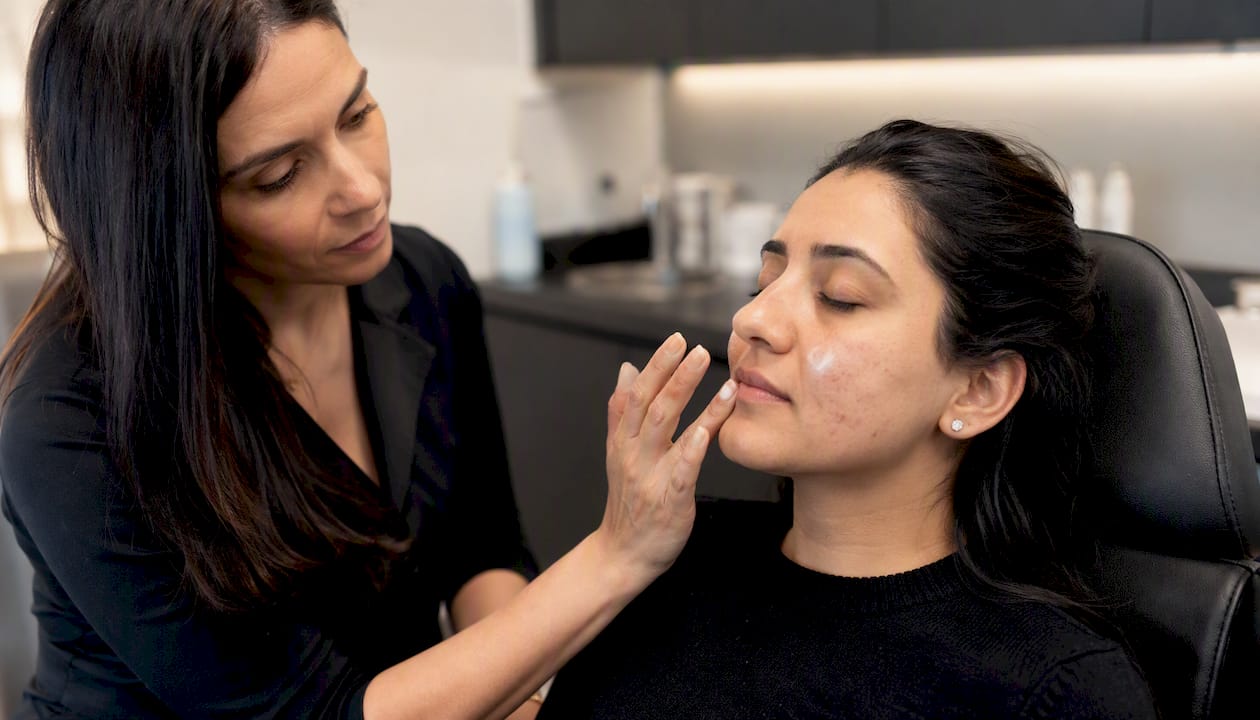
Step 1: Control the underlying inflammation. Treat active acne, eczema, or whatever triggered the PIH in the first place. No lightening regimen works well against an ongoing inflammatory signal.
Step 2: Establish photoprotection. Daily broad-spectrum sunscreen is non-negotiable. UV exposure stimulates melanocytes and undoes weeks of topical therapy in a single afternoon.
Step 3: Start topical lightening agents. Monotherapy works, but combination therapy (a tyrosinase inhibitor plus a retinoid, with or without a short-term topical steroid) produces better outcomes than any single agent.
Step 4: Add procedural options for recalcitrant cases. Chemical peels, lasers, and microneedling are reserved for PIH that does not respond to topicals after several months of consistent use.
Realistic expectations:
- Visible improvement typically takes several weeks of consistent topical use before it is noticeable
- Epidermal PIH often improves over several months
- Dermal PIH may require a prolonged period, and some cases do not fully resolve
- Combination therapy consistently outperforms monotherapy in clinical practice
Topical treatments for PIH: what works and how to use them
Topical therapy is the first-line approach for most cases of PIH. The agents below work through different mechanisms, and combining them is usually more effective than using any one alone.
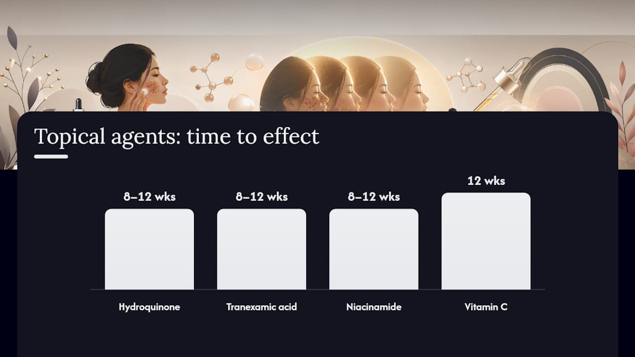
| Agent | Mechanism | Typical concentration | Time to effect | Common side effects |
|---|---|---|---|---|
| Hydroquinone | Tyrosinase inhibitor | 2% (OTC), 4% (Rx) | 8–12 weeks | Irritation, ochronosis (rare, prolonged use) |
| Tretinoin | Increases epidermal turnover | commonly available prescription concentrations | several weeks to months | Dryness, peeling, photosensitivity |
| Azelaic acid | Tyrosinase inhibitor, anti-inflammatory | typical commercial concentrations | several weeks to months | Mild stinging, redness |
| Tranexamic acid | Inhibits melanocyte activation | 2%–5% (topical) | 8–12 weeks | Generally well tolerated |
| Niacinamide | Blocks melanosome transfer | 4%–10% | 8–12 weeks | Minimal |
| Kojic acid | Tyrosinase inhibitor | used in various concentrations | several weeks to months | Contact sensitization possible |
| Vitamin C (ascorbic acid) | Antioxidant, reduces oxidation of melanin | 10%–20% | 12 weeks | Oxidizes quickly; formulation matters |
The steroid component reduces irritation and inflammation but is recommended for short-term use to minimize risks such as skin atrophy and steroid-induced pigment changes.
For acne-associated PIH specifically, the JAAD Delphi consensus supports early acne control with a retinoid plus benzoyl peroxide as the foundation, with lightening agents added as adjuncts when needed.
Safety notes: Patch-test any new agent on the inner arm before applying to the face. Stop any topical if you experience severe burning, blistering, or worsening pigment. Topical steroids beyond 8 weeks carry real risks of atrophy, telangiectasia, and paradoxical pigment changes.
Procedural treatments: when they help and when they backfire
Procedures are not a shortcut for PIH. They are a second-line option for cases that have not responded adequately to topical therapy, and they carry a real risk of making pigment worse if used incorrectly.
Common procedural options:
- Superficial chemical peels (glycolic acid 20–70%, salicylic acid 20–30%): best for epidermal PIH; multiple sessions needed
- Medium-depth peels (trichloroacetic acid 15–35%): more effective but higher risk in darker skin types
- Q-switched Nd:YAG laser: targets melanin with minimal thermal damage; a common choice for skin of color
- Picosecond lasers: deliver energy in shorter pulses, reducing heat and lowering paradoxical PIH risk
- Fractional non-ablative devices: stimulate remodeling with lower downtime than ablative options
- Microneedling and mesoneedling: create controlled micro-injury to stimulate turnover; can be combined with topical agents for enhanced delivery
Priming the skin with a topical lightening agent for 4–6 weeks before any procedure reduces the risk of post-procedure PIH in high-risk patients. This step is frequently skipped in non-specialist settings, which is one of the main reasons procedures go wrong.
Aggressive lasers and peels on unprimed or actively inflamed skin in darker phototypes can trigger paradoxical PIH that is darker and more persistent than the original spots. Experienced clinicians use conservative energy settings, pre-procedure priming, and incremental protocols to protect reactive skin.
Pro Tip: Before booking any laser or peel for PIH, ask the provider specifically about their experience treating your skin phototype, what priming protocol they use, and what energy settings they plan to start with. A provider who cannot answer those questions clearly is not the right provider for this treatment.
For aftercare guidance following any cosmetic procedure, Rao Dermatology’s cosmetic procedure aftercare steps outline the key steps to reduce post-procedure inflammation and protect results.
Daily habits that prevent PIH and help existing spots fade
Prevention is not a passive strategy. These steps actively reduce both the risk of new PIH and the duration of existing spots.
- Apply broad-spectrum SPF 30–60 every morning, including cloudy days and days spent mostly indoors near windows
- Reapply sunscreen every two hours when outdoors, or after sweating and swimming
- Wear sun-protective clothing (UPF 50+ fabric, wide-brim hat) during peak UV hours (10 AM–4 PM)
- Stop picking, squeezing, or scratching any active lesion, no matter how tempting
- Use a gentle, fragrance-free cleanser and avoid abrasive scrubs or rough washcloths on affected areas
- Discontinue irritating products (high-strength acids, physical exfoliants) while treating active PIH
- Follow prescribed acne or eczema regimens consistently to keep the inflammatory trigger under control
- Consider a tinted mineral sunscreen if you have darker skin. Tinted formulas containing iron oxides block visible light, which can stimulate melanocytes independently of UV, a factor standard clear sunscreens miss
Photoprotection is foundational to PIH management and prevents both worsening of existing spots and recurrence after treatment. A daily hyperpigmentation prevention plan that combines sunscreen with gentle skin care gives topical treatments the best possible environment to work.
Pro Tip: Tinted sunscreens with iron oxides are not just cosmetically useful. They provide meaningful protection against visible light, which can drive melanocyte activity in skin of color even when UV exposure is minimal.
How long does PIH take to clear, and what slows it down?
Epidermal PIH typically improves within 6–12 months with consistent photoprotection and topical treatment. Dermal PIH moves much more slowly and may be permanent in some cases.
Factors that prolong PIH:
- Deeper pigment (dermal involvement)
- Ongoing inflammation from untreated acne or eczema
- Continued UV or visible light exposure without adequate protection
- Post-treatment irritation that triggers new inflammation
- Darker skin phototype (more reactive melanocytes)
- Inconsistent or interrupted treatment
Rough milestones:
- Weeks 4–8: Inflammation subsides; topical agents begin working
- Months 2–4: First visible lightening in epidermal PIH with consistent use
- Months 6–12: Significant improvement or near-clearance of epidermal PIH
- Year 1 and beyond: Dermal PIH continues to fade slowly; some cases plateau
Recurrences are common without ongoing photoprotection, even after successful treatment. Maintenance sunscreen use is not optional once spots have cleared.
Treatments to avoid and warning signs that need medical attention
Some of the most common mistakes people make with PIH actually make it worse, sometimes permanently.
Common harmful approaches:
- Prolonged topical steroid use without medical supervision leads to skin thinning, telangiectasia, and steroid-induced dyspigmentation
- Aggressive at-home peels (high-concentration glycolic or TCA without professional guidance) can trigger severe inflammation and paradoxical PIH
- Unverified cosmetic procedures from providers without experience in skin of color carry a high risk of worsening pigment
- Persistent picking or squeezing re-injures skin and restarts the inflammatory cycle
Red flags that warrant prompt evaluation:
- Pigment that is spreading rapidly or changing shape
- Irregular, asymmetric borders that do not match a prior lesion site
- Raised, nodular, or textured areas within the pigmented patch
- Pigment changes in mucous membranes (lips, mouth, gums)
- Systemic symptoms alongside new pigment changes (fatigue, weight change)
- No improvement after four months of consistent, appropriate treatment
Pro Tip: If you have been using a topical steroid on a dark spot for more than 8 weeks without medical guidance, stop and see a dermatologist before continuing. Steroid misuse is one of the most preventable causes of treatment-resistant pigment changes.
How Rao Dermatology evaluates and manages PIH in clinic
At Rao Dermatology, a PIH evaluation follows a structured workflow designed to avoid the most common treatment errors: starting lightening agents before inflammation is controlled, skipping photoprotection counseling, and rushing to procedures before topicals have had a fair trial.
Typical clinic workflow:
- Detailed intake history covering the triggering event, timeline, prior treatments, and current skin care routine
- Focused physical exam with Wood lamp assessment when depth is uncertain
- Identification and treatment of any active inflammatory condition (acne, eczema) before lightening therapy begins
- Individualized topical regimen based on skin phototype, pigment depth, and tolerance
- Photoprotection counseling, including tinted sunscreen recommendations for skin of color
- Baseline photography to track progress objectively at follow-up visits
- Follow-up at 8–12 weeks to assess response and adjust the regimen
Consistent topical regimens and photoprotection drive the best outcomes and reduce the risk of treatment-induced flares. There is no quick fix for PIH, but there is a reliable path forward when the steps are followed in the right order.
What patients should bring to their first appointment:
- Photos of the affected area taken over time, if available
- A list of all current medications and topical products
- Notes on any prior treatments and their results
The psychosocial weight of PIH is real, and so are the options
PIH does not just affect how skin looks. For many patients, it affects confidence, daily routines, and how they feel walking into a room. At Rao Dermatology, that reality shapes how we approach every evaluation: with patience, clear timelines, and a treatment plan built around your skin specifically, not a generic protocol. The spots that feel permanent rarely are, and the path to improvement is more predictable than most patients expect when they first come in.
Rao Dermatology offers personalized PIH evaluation and treatment
Persistent dark spots deserve more than a generic cream recommendation. Rao Dermatology’s medical and cosmetic dermatology services cover the full range of PIH care: prescription topical regimens, photoprotection counseling, procedural options including chemical peels and laser treatments, and esthetic services for adjunctive skin care support.

At your first appointment, a dermatologist will take a complete history, assess pigment depth, identify any active inflammatory triggers, and build a stepwise plan matched to your skin phototype and goals. Bring photos of the affected area over time and a list of products you are currently using. Rao Dermatology serves patients across California, New Jersey, and New York. To schedule a PIH evaluation, visit Raodermatology or contact the location nearest you.
Sources
- Postinflammatory hyperpigmentation - StatPearls - NCBI Bookshelf
- Melanin Hyperpigmentation Disorders, Management: Global 2025 Guideline Summary - Medscape
- Treatment recommendations for acne-associated hyperpigmentation: Results of the Delphi consensus process - JAAD
This article is general information, not a substitute for advice from a qualified doctor. Consult a qualified healthcare professional about your own circumstances before acting on anything here.
FAQ
What causes post-inflammatory hyperpigmentation?
PIH is caused by excess melanin produced after skin inflammation or injury. Common triggers include acne, eczema, burns, insect bites, razor bumps, and certain cosmetic procedures.
How is PIH different from melasma?
PIH follows a specific inflammatory event (a pimple, rash, or wound), while melasma is driven by hormonal factors and UV exposure without a preceding injury. Melasma is typically symmetric and appears on the cheeks, forehead, and upper lip.
How long does post-inflammatory hyperpigmentation last?
Epidermal PIH often improves within 6–12 months with consistent sunscreen use and topical treatment. Dermal PIH, which appears blue-gray, can take years and may not fully resolve.
Can PIH be treated at home?
Mild epidermal PIH can improve with OTC agents like niacinamide, vitamin C, and azelaic acid combined with daily sunscreen. Persistent or deeper pigment benefits from prescription-strength topicals and professional evaluation.
When should I see a dermatologist for dark spots?
See a dermatologist if spots are spreading rapidly, have irregular borders, have not improved after three to four months of consistent care, or if you are unsure of the diagnosis. Rao Dermatology offers structured evaluations to identify the cause and build a targeted treatment plan.
Recommended
Filed under: