
Learn to identify and treat keratosis on legs effectively. Explore types, symptoms, and the best treatment options for healthy skin.
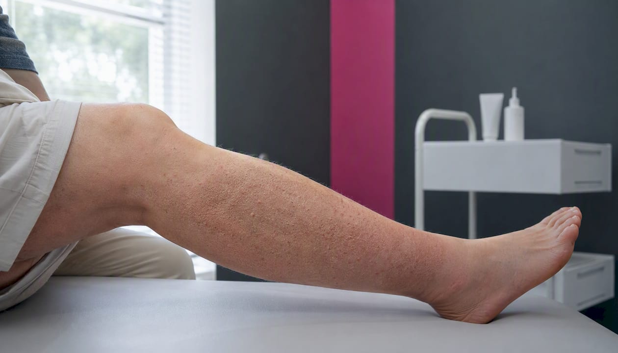
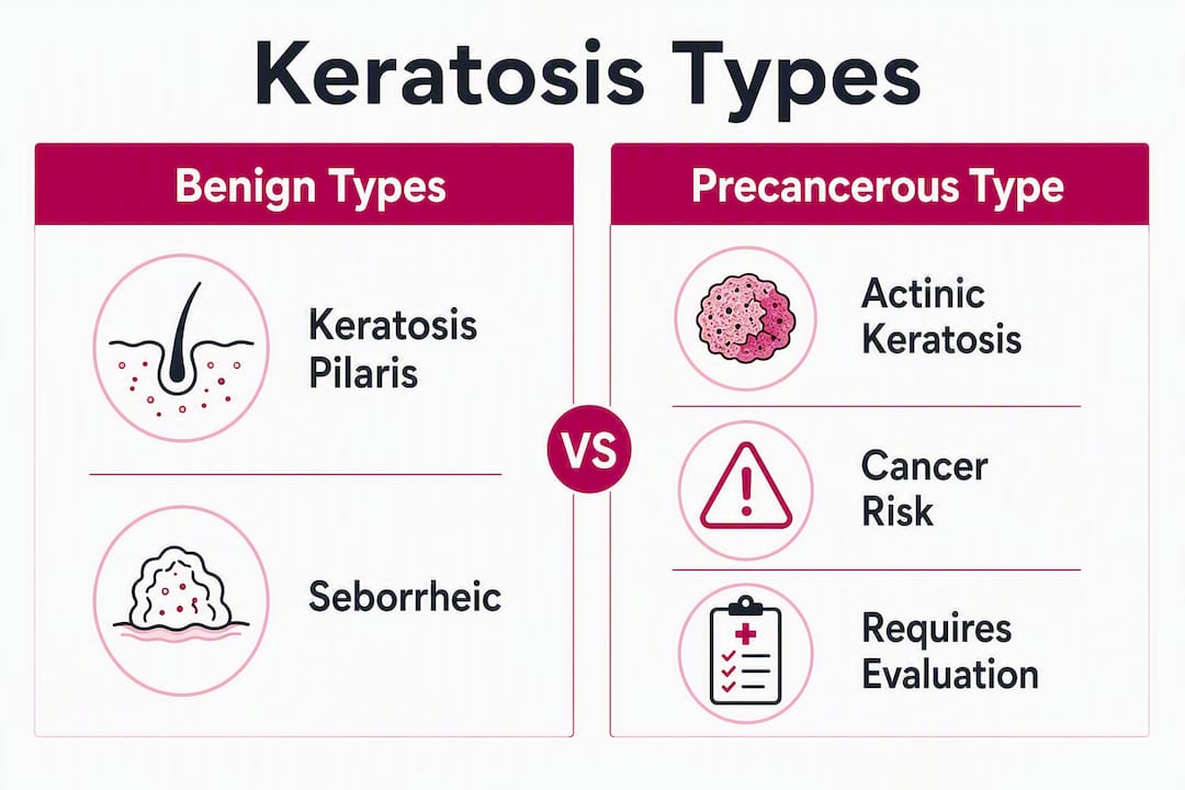
Three types of keratosis account for most of what people notice on their legs: keratosis pilaris (KP), seborrheic keratosis (SK), and actinic keratosis (AK). KP is genetic and benign, showing up as rough, sandpaper-like bumps on the thighs. SK produces waxy, stuck-on brown or black growths that become more common after age 50. AK is the one that demands attention: it’s a precancerous lesion caused by cumulative sun damage that can progress to squamous cell carcinoma if left untreated. For KP, consistent moisturizing and keratolytic products are your main tools. For SK, removal is optional unless a lesion is irritated or changing. For anything that bleeds, grows quickly, crusts, or changes color, skip the self-care aisle and call a dermatologist.
- KP: Benign, genetic, mainly cosmetic. Managed with emollients and keratolytics. No cure, but real improvement is achievable.
- SK: Benign, age-related. Removal is elective unless the lesion is bothersome or changing.
- AK: Precancerous. Requires professional evaluation, possible biopsy, and treatment. The American Academy of Dermatology recommends prompt assessment for any rough, scaly, or discolored patch on chronically sun-exposed skin.
If a lesion is new, changing, bleeding, or non-healing, see a dermatologist rather than waiting.
Table of Contents
- How to tell the three types apart on your legs
- Keratosis pilaris on the legs: what causes it and how to manage it
- Actinic keratosis on the legs: cancer risk, appearance, and treatment
- Seborrheic keratosis on the legs: what to expect and when to remove it
- How dermatologists diagnose keratoses and the red flags that need urgent attention
- Comparing treatment options: what works, what heals, and what to watch for
- Daily skin care and prevention strategies that actually reduce keratosis symptoms
- How Raodermatology evaluates and treats keratoses on the legs
- Key Takeaways
- What dermatologists actually weigh when treating leg keratoses
- Raodermatology offers professional evaluation for keratoses on the legs
- Selected reliable sources for further reading
- FAQ
How to tell the three types apart on your legs
Getting the diagnosis right before you treat anything matters more than most people realize. Here’s how the three types look and feel on the legs.
Keratosis pilaris clusters on the front and outer thighs as tiny, follicular bumps. Run your hand across the skin and it feels like fine sandpaper or a plucked chicken. The bumps are usually skin-colored or faintly red, rarely painful, and don’t change much over time.

Seborrheic keratosis looks like something was stuck onto the skin rather than growing from within it. The surface is waxy or velvety, the color ranges from tan to dark brown or black, and the edges are well-defined. SK can appear anywhere on the legs and tends to multiply with age.
Actinic keratosis feels rougher than it looks. The patch may be only slightly raised, but it has a gritty, scaly texture and can be tender when rubbed. Color varies from skin-toned to pink, red, or brown. On the legs, AK typically appears on areas with the most sun exposure, such as the shins and calves.
- Texture: KP = fine, follicular roughness; SK = waxy and raised; AK = coarse, scaly, sometimes tender
- Color: KP = skin-toned or faintly red; SK = tan to black; AK = variable, often with irregular pigment
- Edges: KP = diffuse; SK = sharply defined; AK = irregular, poorly defined
- Behavior: KP stays stable; SK grows slowly; AK can bleed with minor trauma or crust over
Pro Tip: Color is the least reliable cue, especially on deeper skin tones. Texture and change over time are more useful. A lesion that bleeds when you scratch it or looks different than it did six months ago warrants a dermatology visit regardless of color.
Keratosis pilaris on the legs: what causes it and how to manage it
KP forms when excess keratin blocks individual hair follicles, creating the characteristic rough bumps. It’s genetic, runs in families, and often appears alongside atopic dermatitis or ichthyosis vulgaris. On the legs, the anterior thighs are the most common site. Cold, dry weather tends to worsen it; the bumps often soften in summer or after a warm shower.
The natural history is reassuring. KP is common in children and teenagers and typically resolves by around age 30 in many patients, though some adults carry it indefinitely. There’s no cure, and anyone promising otherwise is overstating the evidence. The realistic goal is durable improvement through a consistent routine, not elimination.
What actually works: twice-daily moisturizing with an emollient-rich cream, combined with a keratolytic ingredient. Urea (10–20%), lactic acid (5–12%), and salicylic acid are the three most evidence-supported options. NHS guidance recommends mild cleansers, regular moisturizing, and gentle exfoliation. Harsh scrubbing or abrasive loofahs tend to inflame the follicles and make things worse.
When to bring a clinician in: if the redness is significant, if the skin is swollen or painful, or if you suspect an overlapping condition like eczema, a dermatologist can sort out what’s driving it and adjust the approach. KP can coexist with atopic dermatitis, and treating only one while missing the other leads to incomplete results.
Actinic keratosis on the legs: cancer risk, appearance, and treatment
Actinic keratosis is a precancerous lesion caused by cumulative UV damage to skin cells. Left untreated, some AKs progress to squamous cell carcinoma. That’s the core reason it gets its own category: it’s not just a cosmetic nuisance.
On the legs, AK typically appears on the shins and calves, areas that accumulate decades of sun exposure. Fair skin, a history of outdoor work or recreation, and a personal or family history of skin cancer all raise the risk. The lesion feels rough and scaly, may be slightly raised, and can bleed with minor friction. Some patients describe a burning or stinging sensation.
Treatment options for AK on the legs include:
- Cryotherapy: Liquid nitrogen freezes the lesion. Fast, office-based, and effective for isolated AKs. Healing on the legs takes longer than on the face, often six weeks or more.
- Topical field therapies: Fluorouracil (5-FU) cream, imiquimod, or diclofenac gel treat both visible lesions and subclinical sun damage across a broader area. Useful when multiple AKs are present.
- Photodynamic therapy (PDT): A photosensitizing agent is applied and activated with a specific light wavelength. PDT is particularly well-suited to field-based treatment of AK, managing subclinical damage while limiting the number of destructive procedures needed.
- Excision or shave biopsy: Reserved for lesions suspicious for progression to squamous cell carcinoma. Provides tissue for pathology review.
Healing time on the legs is a real clinical consideration. Cryotherapy on the legs often requires six weeks or longer to heal, compared to roughly 5–10 days on the face and 3–4 weeks on the hands. Ask your dermatologist about hypopigmentation and scarring risk before committing to a destructive approach, particularly if you have darker skin.
Seborrheic keratosis on the legs: what to expect and when to remove it
SK are benign, waxy growths that become more frequent with age, most commonly in adults over 50. They’re harmless in the strict medical sense: they don’t become cancerous. But they can itch, catch on clothing, or simply bother you cosmetically, and those are legitimate reasons to have them removed.
The stuck-on appearance is the giveaway. SK looks like a brown or black wafer pressed onto the skin surface rather than growing from within it. On the legs, they can appear anywhere and tend to multiply over time.
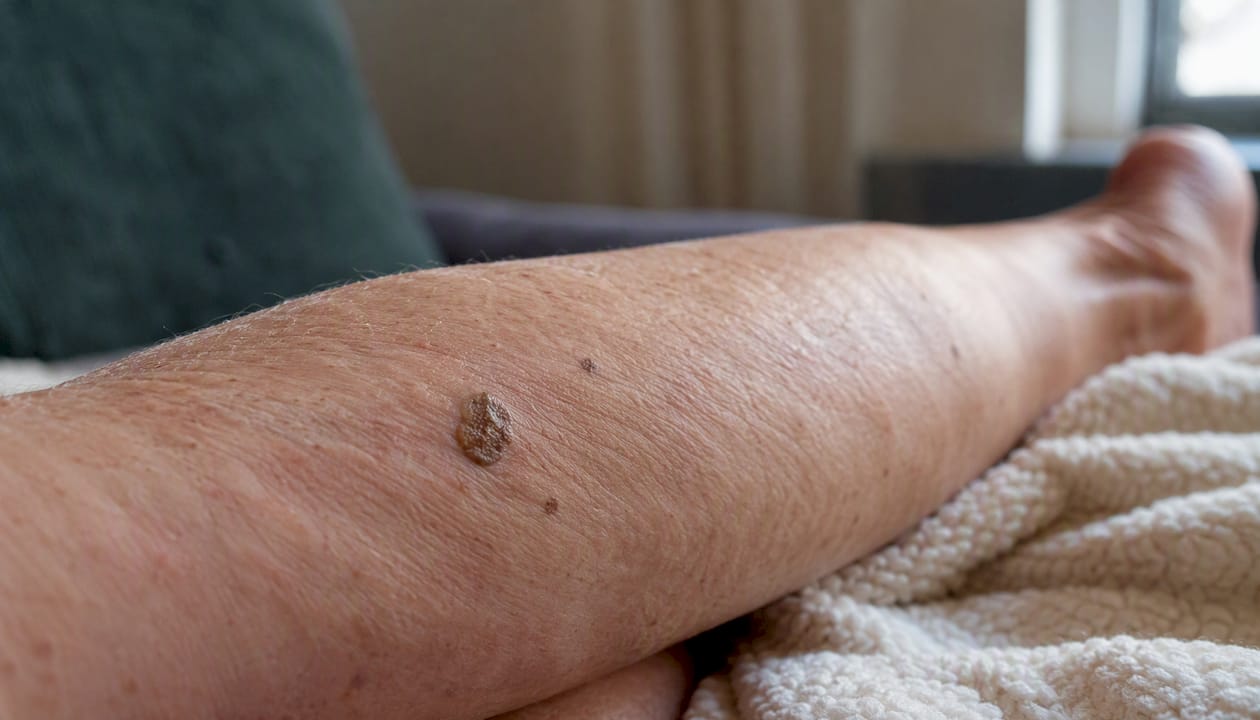
Removal is medically unnecessary unless a lesion is irritated, bleeding, or changing. When removal is the right call, the main options are cryotherapy, curettage, or shave removal. All are effective. The trade-off worth discussing before you proceed: cryotherapy and curettage can cause permanent hypopigmentation, especially in darker skin tones, and the pale patch left behind can be more noticeable than the original growth. On the legs, where healing is slower, that risk is higher than on the face.
One practical rule: don’t attempt DIY removal. Benign-appearing lesions can mimic melanoma or other skin cancers, and self-removal can obscure a cancer diagnosis while also risking infection. A dermatologist can confirm the diagnosis with dermoscopy before any procedure and choose the approach least likely to leave a scar.
How dermatologists diagnose keratoses and the red flags that need urgent attention
Most keratoses are diagnosed clinically: a focused history, a visual exam, and dermoscopy. Dermoscopy lets a clinician inspect surface features, follicular plugs, and pigment patterns that aren’t visible to the naked eye, which is often enough to distinguish KP, SK, and AK with confidence. Biopsy is reserved for lesions that are atypical or suspicious for malignancy, and for AK, dermatopathology review secures the diagnosis when clinical uncertainty remains.
Red flags that warrant prompt evaluation:
- Rapid growth over weeks or months
- Persistent bleeding or crusting without trauma
- New or expanding areas of dark, irregular pigment
- Non-healing ulceration
- Persistent pain or tenderness in a previously stable lesion
Any one of these changes a “watch and wait” approach into a “see a dermatologist this week” situation.
Healing time by treatment site:
| Body site | Typical healing time after cryotherapy |
|---|---|
| Face | 5–10 days |
| Hands | 3–4 weeks |
| Legs | 6 weeks or longer |
These differences matter for treatment planning. A patient with multiple AKs on the shins who needs to stay mobile may be better served by a field therapy than by repeated cryotherapy sessions with prolonged wound care.
Comparing treatment options: what works, what heals, and what to watch for
No single treatment fits every keratosis type, and the legs add their own wrinkle because healing is slower there than almost anywhere else on the body.
| Treatment | Best for | Healing time (legs) | Key trade-offs |
|---|---|---|---|
| Emollients + keratolytics | KP | Ongoing maintenance | No cure; requires consistency |
| Cryotherapy | AK, SK | 6 weeks or longer | Hypopigmentation, scarring risk |
| Topical field therapy (5-FU, imiquimod) | AK (multiple lesions) | 3–4 weeks | Inflammation during treatment |
| Photodynamic therapy | AK (field treatment) | 2–4 weeks | Requires clinic visit; photosensitivity |
| Curettage / shave removal | SK | 4 weeks or longer | Pigment risk in darker skin tones |
| Excision / biopsy | Suspicious AK or SK | 4 weeks or longer | Scar; provides pathology |
A few points worth raising with your dermatologist before committing to a procedure:
- Pigment risk: Cryotherapy and curettage carry a real risk of permanent hypopigmentation, particularly on the legs and in patients with Fitzpatrick skin types IV–VI. Ask specifically about this before treatment.
- Field vs. spot treatment: For patients with multiple AKs, staged field-based approaches reduce the total number of destructive procedures while addressing subclinical damage.
- Insurance coverage: Medically necessary AK treatment is generally covered; cosmetic SK removal typically is not.
Pro Tip: Photograph any lesion you’re monitoring before your appointment. A side-by-side comparison from three to six months earlier gives your dermatologist far more useful information than a verbal description of “it looks bigger.”
Avoid home excision, freezing sprays bought online, or any unlicensed removal method. Beyond infection risk, removing a lesion at home destroys the tissue needed for pathology if the growth turns out to be cancerous.
Daily skin care and prevention strategies that actually reduce keratosis symptoms
Managing keratosis on the legs long-term comes down to two parallel goals: controlling existing symptoms and preventing new sun damage.
For KP and general skin health:
- Cleanse with a gentle, fragrance-free wash. Harsh soaps strip the skin barrier and worsen follicular plugging.
- Apply an emollient immediately after bathing while skin is still slightly damp. Creams outperform lotions for dry, rough skin.
- Use a keratolytic ingredient consistently: urea 10–20%, lactic acid 5–12%, or salicylic acid formulations. These dissolve the keratin plugs rather than just masking them. If you’re unsure which concentration suits your skin, a pharmacist or dermatologist can guide you. For those managing dry or eczema-prone skin alongside KP, gentle cleansing and moisturizing strategies can help protect the skin barrier without aggravating either condition.
- Avoid abrasive scrubs. Physical exfoliation on already-inflamed follicles tends to cause more redness, not less.
For AK prevention:
- Apply broad-spectrum SPF 30+ sunscreen to the legs every day you’ll have sun exposure, not just at the beach. The shins and calves accumulate UV damage year-round.
- Wear UPF-rated clothing when spending extended time outdoors.
- Limit peak sun exposure between 10 AM and 4 PM, especially in summer months.
- Schedule annual skin checks if you have a history of AK, fair skin, or significant lifetime sun exposure.
After a procedure:
Follow your dermatologist’s post-procedure instructions precisely. Sun avoidance while healing reduces hyperpigmentation risk, and proper wound care lowers the chance of infection. Post-treatment skin care on the legs requires more patience than on the face: six weeks is a realistic minimum for full healing after cryotherapy.
How Raodermatology evaluates and treats keratoses on the legs
Raodermatology offers the full range of services relevant to keratosis on the legs: medical dermatology, skin cancer screening and treatment, dermoscopy, dermatopathology, photodynamic therapy, and procedural removal options including cryotherapy, curettage, and excision.
At an appointment, you can expect a focused skin history, a full visual exam, and dermoscopy for any lesion that warrants closer inspection. If a biopsy is indicated, the tissue goes to Raodermatology’s dermatopathology service for interpretation. Treatment planning includes a frank discussion of trade-offs: healing time, pigment risk, number of sessions, and what to expect cosmetically. Aftercare instructions are specific to the procedure and the patient’s skin type.
Raodermatology operates across California, New Jersey, and New York, with 25+ years of experience in both medical and cosmetic dermatology. For scheduling and insurance information, the services page covers the full offering.
Key Takeaways
Actinic keratosis is the only common leg keratosis that carries cancer risk, making professional evaluation non-negotiable for any rough, scaly, or changing lesion on sun-exposed skin.
| Point | Details |
|---|---|
| KP is benign and manageable | Use urea, lactic acid, or salicylic acid consistently; improvement is realistic, cure is not. |
| SK is benign but should be checked if changing | Any SK that bleeds, grows rapidly, or changes color warrants dermoscopy or biopsy. |
| AK is precancerous and requires treatment | AK can progress to squamous cell carcinoma; cryotherapy, topical therapy, or PDT are standard options. |
| Leg healing takes longer | Expect six weeks or more after cryotherapy on the legs; discuss hypopigmentation risk before any procedure. |
| Raodermatology offers full evaluation | Skin cancer screening, dermoscopy, dermatopathology, and PDT are all available across CA, NJ, and NY locations. |
What dermatologists actually weigh when treating leg keratoses
The conversation most patients don’t get before a procedure is the one about trade-offs. Removing a seborrheic keratosis from the shin sounds simple until you explain that the pale patch left by cryotherapy may take months to fade, and in some skin tones, it never fully does. That’s not a reason to avoid treatment. It’s a reason to have the conversation first.
For KP, the clinical instinct to “do more” is usually wrong. Lasers and aggressive procedures are rarely the right first-line approach. The evidence points to patient education, daily keratolytics, and realistic expectations. Most patients who’ve tried every product on the market haven’t tried them consistently enough or at the right concentration. Urea 20% applied twice daily for three months looks different from urea 5% used occasionally.
The AK picture is where I’d push back against a “wait and see” approach most firmly. Patients often assume that because a patch has been there for years without changing, it’s fine. But subclinical sun damage accumulates silently, and the lesion you’ve had for five years is not necessarily safer than one that appeared last month. Annual skin checks for anyone with a history of AK or significant sun exposure aren’t overcautious. They’re the standard of care.
If you’re uncertain about a lesion on your legs, the right move is a dermatology evaluation. Early assessment almost always leads to simpler, less invasive treatment.
Raodermatology offers professional evaluation for keratoses on the legs
Raodermatology’s medical dermatology team evaluates and treats all three types of keratosis on the legs, from straightforward KP management to skin cancer screening and photodynamic therapy for actinic keratosis. The practice brings 25+ years of dermatology experience across California, New Jersey, and New York, with in-house dermatopathology for cases that need biopsy interpretation.

If you have a rough, scaly, or changing lesion on your legs, or simply want a professional baseline skin check, booking an evaluation at Raodermatology is the clearest next step. The services page lists all available treatments and locations. For patients specifically concerned about precancerous lesions, the skin cancer services page outlines the full diagnostic and treatment pathway.
Selected reliable sources for further reading
- Keratosis pilaris — Harvard Health
- Keratosis pilaris — NHS
- Actinic keratosis — Yale Medicine
- Seborrheic keratosis — Cleveland Clinic
- Seborrheic keratosis — MedlinePlus
- Photodynamic therapy for actinic keratosis — Raodermatology
- Skin cancer services — Raodermatology
FAQ
How do you get rid of keratosis bumps on the legs?
For keratosis pilaris, consistent use of keratolytic moisturizers containing urea, lactic acid, or salicylic acid reduces bumps significantly over weeks to months. For actinic keratosis or seborrheic keratosis, a dermatologist can perform cryotherapy, curettage, or photodynamic therapy depending on the type and severity.
What vitamin deficiency causes keratosis?
Vitamin A deficiency is associated with follicular hyperkeratosis, a condition that can resemble KP. However, most cases of keratosis pilaris in the United States are genetic rather than nutritional; supplementing vitamins without a confirmed deficiency is unlikely to resolve typical KP.
What ointment removes keratosis?
No over-the-counter ointment removes keratosis entirely, but prescription-strength urea (10–20%), tretinoin, or fluorouracil cream can significantly reduce actinic keratosis and improve KP texture. A dermatologist can prescribe the appropriate formulation based on the specific type and location.
Can keratosis on the legs spread to other areas?
KP and SK do not spread in the infectious sense, but new lesions can appear over time as part of the same underlying tendency. AK can develop in new sun-exposed areas as cumulative UV damage accumulates, which is why sun protection and annual skin checks matter for anyone with a history of actinic lesions.
When should a keratosis on the legs be biopsied?
A biopsy is warranted when a lesion bleeds without trauma, grows rapidly, develops irregular pigment, fails to heal, or looks atypical under dermoscopy. Distinguishing AK from early squamous cell carcinoma or melanoma requires tissue analysis, not visual assessment alone.
Recommended
- What is Actinic Keratosis? Symptoms, Causes & Treatment Guide | Rao Dermatology
- Actinic Keratosis: Symptoms, Causes, and Treatment | Rao Dermatology
- What Is a Skin Lesion? Types, Causes, and Treatment | Rao Dermatology
- Photodynamic Therapy for Actinic Keratosis: Advanced Treatment at Rao Dermatology | Rao Dermatology
Filed under: