
Discover what is actinic keratosis, its symptoms, causes, and effective treatments. Learn how to protect your skin from UV damage.

Actinic keratosis (AK) is a precancerous patch of skin caused by years of cumulative UV exposure. It’s not cancer yet, but it can become squamous cell carcinoma if left untreated, which is why dermatologists take it seriously and why you should too.
- AKs most often appear on the face, scalp, ears, lips, backs of the hands, and forearms.
- Many patients feel them before they see them: a rough, sandpaper-like texture on otherwise normal-looking skin.
- They are treatable, with multiple evidence-based options available depending on how many lesions you have.
- A single AK is an early warning sign of broader UV damage across the surrounding skin.
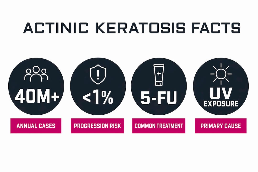
More than 40 million Americans develop actinic keratoses each year. Per-lesion progression risk ranges from less than 1% to about 10% per year, which sounds reassuring until you consider that most patients have multiple lesions at once. If you notice a rough, scaly patch that won’t go away, see a dermatologist.
Table of Contents
- What is actinic keratosis, exactly?
- What do actinic keratoses look and feel like?
- What causes actinic keratosis and who is most at risk?
- What is the cancer risk from actinic keratosis?
- How do doctors diagnose actinic keratosis?
- What are the treatment options for actinic keratosis?
- How do dermatologists decide which treatment to recommend?
- How can you prevent actinic keratosis?
- When should you see a dermatologist?
- Key Takeaways
- The part most articles skip
- Useful sources
- FAQ
What is actinic keratosis, exactly?
Clinically, actinic keratosis is a premalignant epidermal lesion defined by atypical keratinocytes confined to the epidermis, the outermost layer of skin. Chronic UV radiation damages the DNA in these skin cells, causing abnormal growth that hasn’t yet broken through the basement membrane into deeper tissue. That distinction is what separates AK from invasive squamous cell carcinoma, though the histologic continuum between the two is why evidence-based guidelines treat AKs as precursors warranting either treatment or close surveillance.
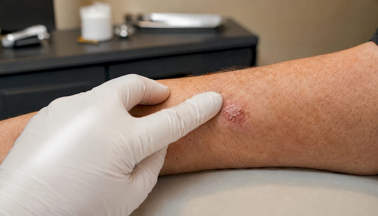
You’ll see several names used interchangeably. Solar keratosis is the most common synonym. Actinic cheilitis refers specifically to AK on the lip vermilion. Senile keratosis is an older term still found in some clinical literature. Histologic variants include hyperkeratotic (thick, wart-like), pigmented (brown or dark), atrophic, and bowenoid forms. Visually, AKs typically appear as rough, scaly, or crusted patches ranging from skin-colored to pink, red, or brownish, usually less than an inch across.
What do actinic keratoses look and feel like?
The tactile cue often comes first. Run your fingertips across sun-exposed skin and an AK feels like a small patch of dried glue or fine sandpaper, even when it barely shows visually. That texture is one of the most reliable early signals.
Visual signs to watch for:
- Rough, scaly, or crusty patch on sun-exposed skin
- Small raised bump or flat area with a dry, flaky surface
- Color ranging from skin-tone to pink, red, or light brown
- A hard, wart-like surface in thicker lesions
- Itching, burning, or tenderness in the area
Common locations:
- Face (especially forehead, nose, cheeks)
- Scalp (particularly in people with thinning hair)
- Ears and lips
- Back of the hands and forearms
- Chest and upper back in people with heavy sun exposure
Pro Tip: Don’t rely on color alone. Run your fingertips across any sun-exposed area during your monthly self-exam. If a spot feels gritty or rough and has been there for more than a few weeks, that texture warrants a professional look, even if it looks unremarkable.
What causes actinic keratosis and who is most at risk?
The mechanism is straightforward: cumulative ultraviolet radiation, both UVA and UVB, damages the DNA in epidermal keratinocytes over time. The damage accumulates across decades. A single bad sunburn doesn’t cause AK; a lifetime of unprotected sun exposure does. Tanning beds deliver the same UV damage and carry the same risk.
Certain people accumulate that damage faster or tolerate it less well:
- Age: Most AKs appear after 40, when decades of UV exposure have built up. Prevalence rises sharply with age.
- Fair skin: People with Fitzpatrick skin types I and II (light skin, light eyes, tendency to burn) have less melanin to absorb UV radiation and are at significantly higher risk.
- History of sunburns or tanning bed use: Each episode adds to cumulative DNA damage.
- Immunosuppression: Organ transplant recipients and others on immunosuppressive medications develop AKs at much higher rates and are at greater risk of progression.
- Occupational or recreational outdoor exposure: Farmers, construction workers, sailors, and lifeguards face disproportionate lifetime UV loads.
- Prior skin cancer: A personal history of squamous cell carcinoma, basal cell carcinoma, or melanoma signals a skin that has already shown susceptibility.
Understanding your own skin cancer risk factors helps put AK in context. The same UV exposure that drives AK development is also the primary driver of all three major skin cancers.

What is the cancer risk from actinic keratosis?
AK is not cancer, but it sits on the continuum toward squamous cell carcinoma (SCC). The honest answer about individual lesion risk is that it’s genuinely uncertain.
| Metric | Estimate | Source |
|---|---|---|
| Americans with AK annually | More than 40 million | AAD |
| Per-lesion annual progression risk | Less than 1% to ~10% | Merck Manual |
| Progression target | Cutaneous squamous cell carcinoma | S3 guideline / StatPearls |
The wide range in progression risk reflects real biological variability. Thicker, hyperkeratotic lesions and those in immunosuppressed patients carry higher risk. The problem is that no current clinical tool reliably predicts which individual lesion will progress. That uncertainty is exactly why dermatologists lean toward treating rather than watching, especially when lesions are multiple or changing.
Long-term prognosis after treatment is generally good, but recurrence is common. New AKs can develop in the same sun-damaged field even after successful treatment of existing lesions. That’s why treatment is paired with ongoing surveillance and sun damage management, not treated as a one-time fix.
How do doctors diagnose actinic keratosis?
Diagnosis is usually clinical. A board-certified dermatologist can identify most AKs through careful visual examination and palpation, sometimes aided by dermoscopy, a handheld device that magnifies the skin surface and reveals vascular and pigment patterns not visible to the naked eye. Most AKs don’t need a biopsy.
Biopsy becomes necessary when a lesion shows features that raise concern for invasive SCC or another diagnosis:
- Rapid growth or change in size, shape, or color
- Spontaneous bleeding or ulceration
- A cutaneous horn (hard, cone-shaped protrusion)
- A non-healing sore or erosion
- Unusual thickness or induration
- Lesions that don’t respond to standard treatment
When a biopsy is performed, the tissue goes to a dermatopathology lab for histologic analysis, which can confirm the degree of dysplasia and rule out invasive carcinoma.
Teledermatology can be a useful first step for patients in remote areas or those triaging whether a spot warrants an in-person visit. That said, dermoscopy and palpation require physical presence, so any lesion with suspicious features should be evaluated in person. The AAD recommends that AKs be diagnosed by a dermatologist, not self-diagnosed, because benign lesions like seborrheic keratoses can look similar but require completely different management.
What are the treatment options for actinic keratosis?
Treatment falls into two broad categories: lesion-directed therapies that target individual spots, and field-directed therapies that treat a broader area of sun-damaged skin. The right choice depends on how many lesions you have, where they are, and your overall health.
Lesion-directed options
- Cryotherapy (liquid nitrogen): The most common in-office procedure. Liquid nitrogen freezes the lesion, which then blisters, crusts, and falls off over 1–2 weeks. Some AKs need a repeat session. Fast, low-cost, and effective for isolated lesions.
- Curettage: The dermatologist scrapes away the lesion with a small curette, sometimes followed by electrodesiccation. Good for thicker or hyperkeratotic lesions.
- Laser resurfacing: Used for larger areas or when cosmetic outcome is a priority, particularly on the face.
Field-directed options
When AKs are scattered across a broad area, treating each spot individually misses the surrounding subclinical damage. Field therapy addresses the whole zone:
- 5-Fluorouracil (5-FU): Applied topically for a 2–4 week regimen; causes significant redness, peeling, and crusting as it works.
- Imiquimod: An immune-response modifier applied over weeks to months (schedules vary from a few weeks to 12–16 weeks depending on the protocol); also causes local inflammation.
- Tirbanibulin: A newer topical approved for a 5-day course, with a milder skin reaction profile than 5-FU.
- Diclofenac gel: Applied over 2–3 months; gentler reaction, useful for patients who can’t tolerate more aggressive topicals.
- Photodynamic therapy (PDT): A photosensitizing agent is applied to the skin, then activated with a specific wavelength of light. PDT typically requires two sessions spaced weeks apart; patients must avoid strong outdoor light for 48 hours after each session. Raodermatology offers PDT for actinic keratosis as part of its advanced treatment options.
Pro Tip: Field therapy is supposed to cause a reaction. Redness, peeling, and crusting during a 5-FU or imiquimod course are signs the medication is working, not signs of a problem. Plan around it: schedule treatment when you can take a week or two away from public-facing commitments, and follow your dermatologist’s instructions for wound care to minimize scarring.
Treatment comparison
| Dimension | Lesion-directed (e.g., cryotherapy) | Field-directed (e.g., 5-FU, PDT) |
|---|---|---|
| Best for | Single or few discrete lesions | Multiple lesions or widespread field damage |
| Mechanism | Physical destruction of individual lesion | Treats subclinical damage across a skin zone |
| Typical duration | 1–2 in-office visits | 5 days (tirbanibulin) to 2–4 months (diclofenac) |
| Common side effects | Blistering, crusting, temporary hypopigmentation | Redness, peeling, crusting, burning |
| Healing time | 1–2 weeks per lesion | 2–6 weeks post-treatment depending on agent |
| Relative effectiveness | High for targeted lesions | Addresses visible and subclinical lesions |
| Cost / insurance | Usually covered; low out-of-pocket | Varies; topicals often covered with prior auth |
How do dermatologists decide which treatment to recommend?
No two patients with AK get the same plan, and that’s by design. Clinical guidelines from the American Academy of Dermatology and the S3 guideline on AK and squamous cell carcinoma both emphasize individualized decision-making based on a cluster of factors.
What clinicians assess:
- Lesion count and distribution: A patient with 2 isolated AKs on the hand gets a different conversation than one with 20 scattered across the scalp. The latter has field cancerization, meaning the surrounding skin carries subclinical UV damage that lesion-directed therapy alone won’t address.
- Lesion thickness and type: Hyperkeratotic or indurated lesions raise more concern and may warrant biopsy before or instead of topical treatment.
- Immune status: Immunosuppressed patients need more aggressive surveillance and often more frequent treatment cycles.
- Age and overall health: Frail older patients may not tolerate prolonged topical regimens; a quick cryotherapy session may be more practical.
- Cosmetic concerns: Lesions on the face, especially in younger patients, may favor treatments with better cosmetic outcomes.
- Patient preference and compliance: A 4-week 5-FU course requires daily application and tolerance of visible skin reaction. If a patient won’t complete it, a different approach is more appropriate.
At your visit, expect the dermatologist to review your history of skin cancer, lifetime sun exposure, current medications (especially immunosuppressants), any prior AK treatments, and your goals for the visit. Bring a list of your medications and be ready to point out any spots you’ve noticed changing.
How can you prevent actinic keratosis?
Prevention doesn’t undo existing AKs, but it meaningfully slows the development of new ones. Consistent sun protection over years reduces the rate at which UV damage accumulates in the skin.
- Apply a broad-spectrum sunscreen with SPF 30 or higher every morning, even on cloudy days, and reapply every two hours during outdoor activity.
- Wear UPF-rated clothing, wide-brim hats, and UV-blocking sunglasses when outdoors for extended periods.
- Seek shade between 10 AM and 4 PM, when UV intensity peaks.
- Avoid indoor tanning entirely. Tanning beds emit concentrated UV radiation with no safe threshold.
- Perform a monthly skin self-exam: use good lighting, check all sun-exposed areas, and pay attention to texture as well as appearance.
Patients who commit to consistent sun protection typically see fewer new AKs over time, though the timeline is measured in years, not weeks. Existing lesions won’t resolve on their own with sunscreen; they need treatment. Prevention is about what comes next.
There are no proven actinic keratosis home remedies that reliably clear established lesions. Over-the-counter products are not substitutes for prescription topicals or in-office procedures. Self-treating a suspicious lesion risks delaying diagnosis of something that has already progressed.
When should you see a dermatologist?
See a dermatologist promptly if any skin lesion:
- Grows rapidly over weeks
- Bleeds without injury
- Develops an ulcer or open sore that won’t heal
- Becomes painful or tender
- Develops a hard, horn-like protrusion
- Changes significantly in color, size, or texture
These are red flags for possible progression to squamous cell carcinoma and warrant evaluation without delay. Raodermatology’s skin cancer screening and treatment services are available across its California, New Jersey, and New York locations for patients who need a thorough evaluation.
For routine follow-up after AK treatment, most dermatologists schedule a check at 3–6 months post-treatment to assess clearance and look for new lesions. Patients with multiple AKs, a history of skin cancer, or immunosuppression typically need more frequent visits, sometimes every 3–4 months. The goal isn’t just treating what’s there today; it’s catching what develops next.
Key Takeaways
Actinic keratosis is a treatable precancerous skin lesion driven by cumulative UV damage, and catching it early is the most reliable way to prevent progression to squamous cell carcinoma.
| Point | Details |
|---|---|
| AK is precancerous, not cancer | Per-lesion progression risk ranges from less than 1% to ~10% per year; treat proactively. |
| Texture is the first clue | Many AKs feel rough like sandpaper before they look like anything; check by touch. |
| Treatment is highly individualized | Lesion count, immune status, and patient preference all shape the treatment plan. |
| Field therapy causes visible reaction | Redness and peeling from 5-FU or imiquimod are expected and signal the medication is working. |
| Prevention slows new lesion development | Daily SPF 30+ sunscreen and protective clothing reduce future UV damage over years. |
The part most articles skip
Most AK content focuses on the lesion in front of you. What gets less attention is the skin around it.
When a dermatologist sees multiple AKs clustered on your scalp or forearm, the concern isn’t just those visible spots. The surrounding skin has absorbed the same decades of UV radiation. It carries subclinical damage that doesn’t show up yet but may surface as new AKs within months of treating the current ones. That’s the concept of field cancerization, and it’s why field-directed therapies exist. Treating only what you can see is like fixing the cracks in a wall without addressing the settling foundation.
The other thing worth saying plainly: the per-lesion cancer risk numbers (less than 1% to ~10% per year) sound small in isolation. But a patient with 15 AKs, each carrying even a 1% annual risk, faces a meaningfully different cumulative picture than those numbers suggest at first glance. Dermatologists aren’t being overcautious when they recommend treatment for lesions that “don’t look that bad.” They’re accounting for the whole field, the whole patient, and the whole trajectory.
Useful sources
- Actinic Keratosis — StatPearls, NCBI Bookshelf
- Actinic Keratosis Overview — American Academy of Dermatology
- Actinic Keratosis: Diagnosis and Treatment — American Academy of Dermatology
- Actinic Keratoses — Merck Manual Professional Edition
- S3 Guideline: Actinic Keratosis and Squamous Cell Carcinoma (AWMF)
- Actinic Keratosis — MedlinePlus Medical Encyclopedia
For diagnosis and personalized treatment, consult a board-certified dermatologist. This article is general health information, not medical advice. Always confirm your specific situation with a qualified clinician.
FAQ
Is actinic keratosis a type of cancer?
No. AK is a precancerous lesion, meaning the abnormal cells are confined to the outer skin layer and have not invaded deeper tissue. Left untreated, some AKs can progress to squamous cell carcinoma.
How do you get rid of actinic keratoses?
Treatment options include cryotherapy, topical medications like 5-fluorouracil, imiquimod, tirbanibulin, or diclofenac gel, and photodynamic therapy. The right choice depends on lesion count, location, and your overall health, so a dermatologist should guide the decision.
How long does it take for an AK to turn into cancer?
There’s no fixed timeline. Per-lesion annual progression risk ranges from less than 1% to about 10%, and most AKs never progress at all. Because individual risk is unpredictable, dermatologists generally recommend treating rather than waiting.
What do actinic keratoses look like?
AKs typically appear as rough, scaly, or crusty patches on sun-exposed skin, ranging in color from skin-tone to pink, red, or light brown. Many patients notice the texture first: a sandpaper-like feel on an area that may look nearly normal.
Recommended
- What is Actinic Keratosis? Symptoms, Causes & Treatment Guide | Rao Dermatology
- Photodynamic Therapy for Actinic Keratosis: Advanced Treatment at Rao Dermatology | Rao Dermatology
- Skin Cancer Prevention, Detection & Treatment | Rao Dermatology
- Atopic Dermatitis: Complete Guide to Symptoms, Causes, and Treatment Options | Rao Dermatology
Filed under: