
Learn what melanoma is, its signs, causes, and effective treatments. Early detection can significantly improve survival rates.

Melanoma is defined as a cancer that originates in melanocytes, the pigment-producing cells found primarily in the skin. It is the most dangerous form of skin cancer because it spreads to other organs faster than any other skin cancer type. Despite accounting for less than 2% of skin cancer cases in the U.S., melanoma causes the majority of skin cancer deaths, with roughly 104,960 new cases and 8,430 estimated deaths expected in 2025. Catching it early changes everything. When melanoma is found while still localized, the five-year survival rate reaches 99.6%.
What is melanoma and where does it come from?
Melanoma is a malignant tumor that develops when melanocytes mutate and begin growing out of control. Melanocytes produce melanin, the pigment that gives skin its color and provides some protection against UV radiation. When UV rays or other triggers damage the DNA inside these cells, normal growth regulation breaks down. The result is a tumor that can invade surrounding tissue and, if untreated, travel through the lymphatic system or bloodstream to distant organs.
Most melanomas appear on the skin, but the disease can also develop in the eyes, known as ocular melanoma, and in mucous membranes lining the mouth, nose, or digestive tract. This matters because patients and even some clinicians focus only on visible skin spots. A melanoma on the retina or inside the nasal cavity carries the same biological threat as one on the back.

The four main melanoma types recognized clinically are superficial spreading melanoma, nodular melanoma, lentigo maligna melanoma, and acral lentiginous melanoma. Superficial spreading is the most common, growing outward across the skin surface before it invades deeper layers. Nodular melanoma is the most aggressive, growing downward quickly. Acral lentiginous melanoma appears on palms, soles, and under nails, and it is the most common type in people with darker skin tones.
What causes melanoma and who is at risk?
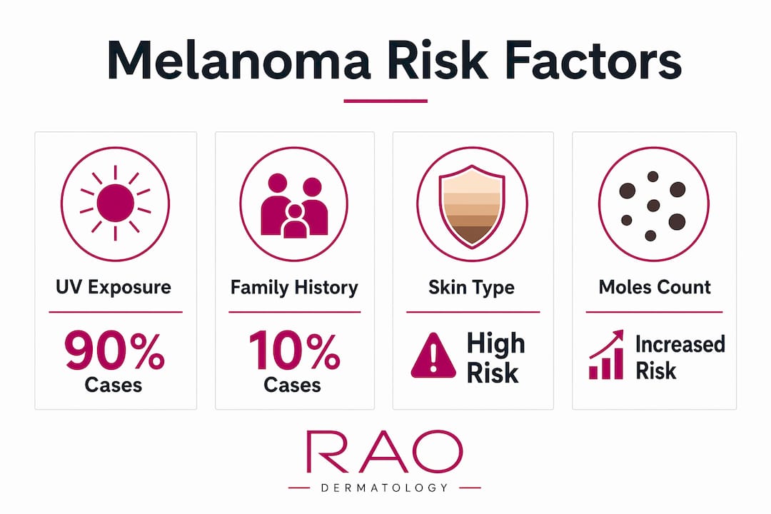
UV radiation is the dominant driver of melanoma. About 93% of melanomas are linked to environmental UV exposure, with blistering sunburns in childhood carrying particular weight. That statistic means sun protection starting in early life is one of the most effective prevention strategies available.
Several factors raise individual risk significantly:
- UV exposure history: Frequent sunburns, especially blistering burns before age 18, increase lifetime risk.
- Family history: A first-degree relative with melanoma raises your risk 6 to 8 times compared to the general population. Understanding your genetic predisposition helps guide screening frequency.
- Skin type and coloring: Fair skin, light eyes, red or blond hair, and a tendency to burn rather than tan all signal higher susceptibility.
- Mole count and type: Having more than 50 common moles, or any atypical moles with irregular borders or mixed colors, raises risk substantially.
- Tanning bed use: Indoor tanning devices emit concentrated UV radiation and are classified as a known carcinogen by the World Health Organization.
- Weakened immune system: Patients on immunosuppressive therapy after organ transplants face elevated risk.
Knowing your personal risk factors is the first step toward a prevention plan that actually fits your situation.
What are the signs and symptoms of melanoma?
The ABCDE rule is the standard clinical framework for identifying suspicious lesions. Dermatologists use it as a first-pass filter during skin exams, and patients can apply it during self-checks.
- A — Asymmetry: One half of the mole does not match the other.
- B — Border: Edges are ragged, notched, or blurred rather than smooth and defined.
- C — Color: Multiple shades of brown, black, red, white, or blue appear within a single lesion.
- D — Diameter: A spot larger than 6 millimeters (roughly the size of a pencil eraser) warrants attention, though melanomas can be smaller.
- E — Evolution: Any change in size, shape, color, or texture over weeks or months is the most important warning sign.
Stability over time in a mole is generally reassuring. A lesion that has looked the same for years carries lower risk than one that changed noticeably in the past few months.
Melanoma does not always follow the ABCDE pattern. Melanoma can develop on the eyes, mucosa, scalp, nails, feet, and inside the mouth. Subungual melanoma, which grows under the nail, often looks like a dark streak running the length of the nail and is frequently mistaken for a bruise or fungal infection.
Pro Tip: Remove nail polish before any skin cancer exam. Nail beds are a critical but easily overlooked site for melanoma, and polish prevents the dermatologist from seeing the full nail surface.
How is melanoma diagnosed and staged?
Diagnosis follows a clear sequence. A dermatologist first performs a full-body skin exam, which typically takes 10 to 15 minutes for a routine check. Patients with many moles or a complex history should expect a longer appointment due to dermoscopy imaging and documentation.
When a lesion looks suspicious, the next step is biopsy. The process generally follows this order:
- Clinical evaluation: The dermatologist examines the lesion with dermoscopy, a magnified light tool that reveals subsurface structure invisible to the naked eye.
- Biopsy selection: Excisional biopsy is preferred because it removes the entire lesion and allows accurate measurement of tumor thickness, called Breslow depth. Thickness is the single most important factor in staging and prognosis.
- Pathology review: A pathologist examines the tissue under a microscope to confirm the diagnosis and assess features like ulceration and mitotic rate.
- Imaging: For thicker tumors or suspicious lymph nodes, CT scans, PET scans, or sentinel lymph node biopsy map whether the cancer has spread.
Staging runs from Stage 0 (melanoma in situ, confined to the outer skin layer) through Stage IV (distant metastasis). Survival rates shift dramatically across stages:
| Stage | Description | 5-Year Survival Rate |
|---|---|---|
| Localized (I/II) | Confined to original site | 99.6% |
| Regional (III) | Spread to nearby lymph nodes | 73.9% |
| Distant (IV) | Spread to other organs | 35.1% |
These survival figures make the case for early detection more clearly than any general warning. The gap between localized and distant disease is not marginal. It is the difference between a near-certain cure and a serious fight.
Pro Tip: Ask your dermatologist to photograph any moles they are monitoring. Serial photography over 6 to 12 months reveals subtle changes that are impossible to track by memory alone.
What are the current treatment options for melanoma?
Treatment depends on stage, tumor location, and the patient’s overall health. Surgery remains the foundation for early-stage disease.
- Wide local excision: The surgeon removes the tumor along with a margin of healthy tissue. For Stage I and II melanomas, this is often the only treatment needed.
- Sentinel lymph node biopsy: Performed alongside excision for tumors thicker than 0.8 millimeters to check whether cancer has reached the lymphatic system.
- Immunotherapy: Immune checkpoint inhibitors, particularly PD-1 blockers like pembrolizumab and nivolumab, have reshaped advanced melanoma care. These drugs release the immune system’s natural brakes, allowing it to attack cancer cells directly.
- Targeted therapy: Patients whose tumors carry a BRAF gene mutation (present in roughly half of all melanomas) can receive BRAF and MEK inhibitor combinations, which block the specific molecular pathway driving tumor growth.
- Radiation therapy: Used selectively for brain metastases or when surgery is not possible.
“Modern treatment advances including immune checkpoint inhibitors have extended median overall survival for advanced melanoma to approximately 6 years, a significant improvement over historical outcomes where patients with Stage IV disease rarely survived beyond 12 months.” Drug Therapy for Melanoma: Current Updates and Future Prospects
That shift in median survival from under one year to roughly six years represents one of oncology’s most dramatic turnarounds in the past two decades. Ongoing clinical trials are testing combinations of immunotherapy and targeted therapy, cancer vaccines, and adoptive cell therapies. For patients diagnosed today, the treatment pipeline is more active than at any prior point in the history of this disease. Learning about advanced melanoma care can help patients ask better questions at their appointments.
Key Takeaways
Melanoma is highly curable when caught early, but it becomes dramatically harder to treat once it spreads beyond the original site.
| Point | Details |
|---|---|
| Melanoma origin | Melanoma starts in melanocytes and can appear on skin, eyes, nails, and mucous membranes. |
| UV exposure is the top cause | About 93% of melanomas link to UV radiation, making sun protection a primary prevention tool. |
| ABCDE rule guides detection | Use Asymmetry, Border, Color, Diameter, and Evolution to evaluate any suspicious spot. |
| Stage determines survival | Localized melanoma carries a 99.6% five-year survival rate; distant spread drops that to 35.1%. |
| Immunotherapy changed outcomes | Checkpoint inhibitors have extended median survival for advanced melanoma to approximately 6 years. |
What I’ve learned from watching patients wait too long
Patients often assume melanoma looks dramatic. They expect something obviously alarming, a large black mass that is clearly wrong. The reality is that many melanomas I have seen in clinical settings look unremarkable at first glance. A slightly irregular mole on the back. A dark streak under a toenail that the patient attributed to an old injury. A flat, pale patch on the scalp hidden under hair.
The most dangerous misconception I encounter is that melanoma only affects people who spent decades at the beach without sunscreen. Family history, mole count, and immune status all matter independently of sun exposure history. A patient with a strong family history and fair skin who has been careful about sun protection still needs regular screening.
Self-exams are valuable, but they have real limits. Patients cannot see their own scalp, back, or the spaces between their toes without help. A partner or a mirror system helps, but nothing replaces a trained eye with a dermoscope. Annual skin cancer screening is the standard recommendation for high-risk patients, and routine skin exams are what make near-total cure rates achievable for early-stage disease.
The patients who do best are not the ones who worried the most. They are the ones who showed up consistently and caught changes early.
— Krunal
Skin cancer screening at Raodermatology
Raodermatology offers comprehensive skin cancer services across its California, New Jersey, and New York locations, backed by 25+ years of clinical experience led by Dr. Babar K. Rao.

The practice combines full-body dermoscopy exams, biopsy services, and personalized prevention planning for patients at every risk level. Whether you have a specific spot that concerns you or want a baseline screening, Raodermatology’s medical dermatology team provides the clinical depth to detect melanoma at its earliest and most treatable stage. Scheduling a skin exam is a straightforward step that can make a meaningful difference in long-term outcomes.
FAQ
What is melanoma in simple terms?
Melanoma is a type of skin cancer that starts in the cells responsible for skin pigment. It is the most dangerous skin cancer because it spreads quickly if not caught early.
How do I detect melanoma at home?
Use the ABCDE rule: check moles for Asymmetry, irregular Borders, multiple Colors, Diameter over 6mm, and Evolution or change over time. Any spot that changes in weeks or months warrants a dermatologist visit.
What causes melanoma to develop?
UV radiation from sunlight or tanning beds causes the majority of melanomas by damaging DNA in melanocytes. Family history of melanoma raises individual risk 6 to 8 times above the general population.
Can melanoma appear somewhere other than the skin?
Yes. Melanoma can develop in the eyes, under the nails, on the scalp, inside the mouth, and on mucous membranes. These sites are easy to miss without a thorough clinical exam.
What melanoma treatment options are available today?
Treatment ranges from surgical excision for early-stage disease to immunotherapy and targeted therapy for advanced cases. Immune checkpoint inhibitors have extended median survival for Stage IV melanoma to approximately 6 years.
Recommended
- Types of skin cancer: Risks, signs, and prevention explained | Rao Dermatology
- What Is a Skin Lesion? Types, Causes, and Treatment | Rao Dermatology
- Skin Cancer Prevention, Detection & Treatment | Rao Dermatology
- Melanoma: A Complete Guide to Skin Cancer Detection, Prevention & Treatment | Rao Dermatology
Filed under: